
Introduction | Welcome to the Microscopic World
Hello students! Welcome back to our biology classroom. Today, we are going to start an incredible journey into a world that is completely invisible to our naked eyes, yet it dictates every single thing about our existence. Have you ever looked at a massive elephant, a towering mango tree, and a tiny ant, and wondered, “Is there a common thread that connects all of us?”
The answer is a resounding yes! Just like millions of individual bricks come together to build a magnificent skyscraper, all living organisms are built from microscopic building blocks. These ultimate units of life are what we call cells. In this chapter, we are going completely microscopic. We are going to tear down the walls of the cell and peek inside to see the machinery that keeps you breathing, thinking, and growing. Whether you are studying a single-celled bacteria that causes a sore throat, or the complex neurons firing in your brain right now, understanding the cell is the absolute fundamental key to unlocking the mysteries of biology. Grab your imaginary microscopes, class, and let’s dive right in!
1. What exactly is a Cell?
When we look at the natural world, we can divide everything into living and non-living. But what gives a living thing its “aliveness”? It is the presence of a cell. Some organisms, like amoebas and bacteria, are entirely made of just one single cell. We call them unicellular. They are the ultimate lone wolves—capable of existing independently and performing every single essential life function (like eating, breathing, and reproducing) all by themselves within that single boundary.
On the other hand, complex beings like you and me are multicellular, made up of trillions of cells working together in perfect harmony. But remember this golden rule: anything less than a complete, intact cell structure cannot sustain independent life. Therefore, the cell is the fundamental structural and functional unit of all living organisms.
Historically, Antonie von Leeuwenhoek was the pioneer who first observed a living cell swimming around under his primitive microscope. Later, the brilliant Robert Brown made another massive breakthrough when he discovered the dense, dark structure inside the cell, which he named the nucleus.
2. The Famous Cell Theory
Pay close attention here, students. The formulation of the Cell Theory is a favorite topic for board examiners!
The story of the cell theory is a fantastic example of scientific teamwork. In 1838, a German botanist named Matthias Schleiden spent his days examining countless plants. He made a bold declaration: all plants are made of various types of cells, which group together to form plant tissues.
A year later, in 1839, a British zoologist named Theodore Schwann was studying animal cells. He noticed that animal cells had a thin, flexible outer boundary (which we now proudly call the plasma membrane). Interestingly, Schwann also peeked at plant cells and noted that they possessed a rigid, extra layer called the cell wall, which animal cells entirely lacked. Putting all this together, Schleiden and Schwann jointly formulated the Cell Theory, stating that all plants and animals are composed of cells and their products.
But there was one glaring mystery they couldn’t solve: Where do these new cells come from? Do they magically pop out of thin air?
Enter Rudolf Virchow in 1855. He observed dividing cells and gave us the legendary Latin phrase: “Omnis cellula-e cellula”. Translated to simple English, this means that all new cells arise only from pre-existing, older cells. Virchow modified the original theory, giving us the modern Cell Theory as we understand it today:
- All living organisms are composed of cells and products of cells.
- All cells arise from pre-existing cells through cell division.
3. An Overview: Size, Shape, and Basic Types
If you scrape the inside of your cheek and place it under a microscope, or peel a thin layer of onion skin, what do you see? You see distinct compartments.
Cells are not a “one-size-fits-all” deal. They exhibit a mind-boggling variety in shape, size, and activity.
- Size: The smallest cells known to mankind are Mycoplasmas, measuring a mere 0.3 micrometers. Bacteria usually hover around 3 to 5 micrometers. Care to guess the largest single isolated cell? It’s the massive egg of an ostrich! In your own body, red blood cells are about 7 micrometers across, while nerve cells can be up to a meter long, making them the longest cells in the human body.
- Shape: Form follows function. A red blood cell is a biconcave disc to squeeze through tight blood vessels. A nerve cell is heavily branched like a tree to pass electrical messages quickly. Some cells are pillar-like (columnar), cubical, or even constantly changing shape like the amoeba.
Prokaryotic vs. Eukaryotic: The Great Divide
Based on the internal architecture, we classify all cells on Earth into two grand categories.
If a cell has a dense, prominent central control room bound by a proper membrane—a true nucleus—we call it Eukaryotic (Eu = true, Karyon = nucleus). These cells also contain specialized, membrane-bound “mini-organs” called organelles.
If a cell is more primitive and lacks this distinct nuclear membrane, leaving its genetic material floating naked in the cell fluid, it is Prokaryotic (Pro = primitive). They do not have complex membrane-bound organelles. However, both types share a jelly-like fluid called cytoplasm, which fills the cell and acts as the main arena where the magic of cellular chemistry happens.
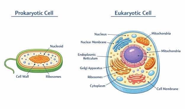
4. The Minimalists: Prokaryotic Cells
Think of a prokaryotic cell like a studio apartment. Everything happens in one open space; there are no separate rooms with doors. This group is represented by bacteria, blue-green algae (cyanobacteria), Mycoplasma, and PPLO. They are generally much smaller and multiply incredibly fast compared to our cells.
4.1 The Cell Envelope
Bacterial cells wear multiple layers of armor to survive harsh environments. This outer covering is a tightly bound three-layered structure:
- Glycocalyx: The outermost sticky layer. If it is loose, we call it a slime layer. If it is thick, tough, and rigidly protective, it is called a capsule.
- Cell Wall: The middle layer. It determines the bacterium’s shape and provides immense structural support, preventing the tiny cell from bursting when water rushes in.
- Plasma Membrane: The innermost layer, semi-permeable just like ours, allowing interaction with the outside world.
Teacher’s Note: You might have heard of “Gram-positive” and “Gram-negative” bacteria. This classification is based entirely on the differences in these cell envelopes and how they respond to a specific chemical stain invented by a scientist named Gram.
4.2 Internal Features and Extensions
Since they don’t have organelles, how do they do complex things like respiration? They have an ingenious solution: the Mesosome. This is a specialized, highly folded inward extension of the plasma membrane. It comes in the form of vesicles, tubules, and lamellae. It massively increases the surface area for enzymes to attach, aiding in respiration, secretion, and even DNA replication!
Genetic Material: They have a single, circular main chromosome floating freely in a region called the nucleoid. Interestingly, many bacteria also have extra, tiny circular DNA rings called Plasmids. These plasmids give the bacteria “superpowers,” like the ability to resist antibiotic medicines!
Surface Structures: If they need to swim, they use long whip-like tails called Flagella (made of a filament, hook, and basal body). If they need to attach to a rock in a stream or to your throat tissues, they use tiny bristle-like fibers called Fimbriae or tubular structures called Pili.
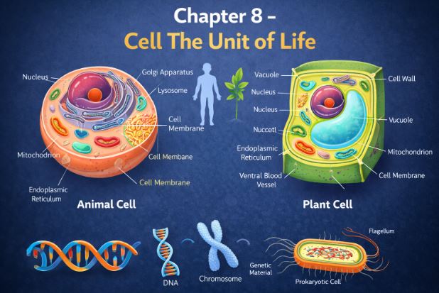
5. The Compartmentalized Mansions: Eukaryotic Cells
Now, let’s step into the complex world of plants, animals, fungi, and protists. Eukaryotic cells are like massive mansions with specialized rooms (organelles) for every specific task.
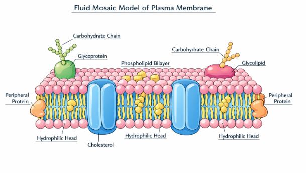
5.1 The Plasma Membrane (The Security Guard)
Every cell needs a boundary that decides what enters and what exits. The most widely accepted model to describe this structure is the Fluid Mosaic Model, proposed by scientists Singer and Nicolson in 1972.
Imagine a vast, oily ocean made of a double layer of lipids (specifically, phospholipids). The lipid molecules have a “water-loving” (polar) head facing the outside and “water-hating” (hydrophobic) tails tucked safely inside, away from the watery cell environment. Floating in this oily lipid ocean are giant protein “icebergs.” Some proteins sit purely on the surface (peripheral), while others are deeply embedded (integral).
Why is it called fluid? Because it is not a rigid wall! The lipid and protein molecules can actually glide and move laterally around each other. This fluidity is crucial for cell division, cell growth, and engulfing food.
Transport: The membrane is selectively permeable. Water and neutral gases can easily slip through the lipid layer without any energy—this is called passive transport (or osmosis, for water). However, if the cell needs to pump something against its natural flow (from low to high concentration), it has to spend energy in the form of ATP. This is called Active Transport (like the famous Sodium-Potassium pump).
5.2 The Cell Wall (The Plant’s Armor)
Plants and fungi cannot run away from danger or bad weather, so they have an extra rigid, non-living armor outside their plasma membrane called the cell wall. It protects them from mechanical damage and infection. In plants, it is made primarily of a tough carbohydrate called cellulose. Interestingly, plant cells are glued together by a sticky middle layer called the middle lamella (made of calcium pectate). To allow neighboring plant cells to communicate and share materials, they have tiny cytoplasmic bridges passing right through the walls, called Plasmodesmata.
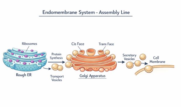
5.3 The Endomembrane System (The Cellular Factory)
Inside the cytoplasm, there are four specific organelles that work together so closely that we group them as the endomembrane system. Their functions are perfectly coordinated, like an assembly line in a factory.
- Endoplasmic Reticulum (ER): A massive network of tiny tubular structures. If it has tiny dot-like ribosomes attached to its surface, it looks bumpy and is called Rough ER (RER). RER is the protein manufacturing unit. If it has no ribosomes, it looks smooth and is called Smooth ER (SER). SER is the factory for making lipids and steroid hormones.
- Golgi Apparatus: Discovered by Camillo Golgi, this looks like a stack of flat, disc-shaped pancakes called cisternae. Think of it as the cell’s post office. Materials (like proteins) coming from the ER enter the receiving side (cis face), get chemically modified and packaged into vesicles, and are shipped out from the shipping side (trans face) to their final destination.
- Lysosomes: These are tiny bubbles pinching off from the Golgi, packed with highly destructive, acidic digestive enzymes. They act as the cell’s waste disposal system, breaking down old organelles, bacteria, or carbohydrates. If the cell is severely damaged, lysosomes burst and digest the entire cell, which is why they are often nicknamed “suicide bags.”
- Vacuoles: A membrane-bound storage sac. In animal cells, they are small and temporary. But in plant cells, a single central vacuole can take up to 90% of the cell’s volume! It stores water, sap, and waste. The membrane covering the plant vacuole is called the Tonoplast.
5.4 The Energy Generators: Mitochondria & Plastids
These organelles are unique because they are not part of the endomembrane system, and they actually contain their own circular DNA and 70S ribosomes! Because of this, they can divide on their own.
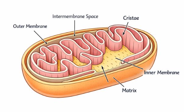
Mitochondria: The undisputed “Powerhouses of the cell.” They are double-membrane bound. The outer membrane is smooth, but the inner membrane is deeply folded inward into structures called Cristae. Why the folds? To massively increase the surface area needed for the enzymes that generate energy (ATP) through aerobic respiration. The jelly inside is called the matrix.
Plastids: Found exclusively in plant cells and some algae. They are large and often contain pigments.
- Chloroplasts: Contain the green pigment chlorophyll. Inside, they have a fluid called stroma and stacks of coin-like flattened sacs called thylakoids. A whole stack of thylakoids is called a granum. This is where solar energy is trapped to cook food (photosynthesis).
- Chromoplasts: Contain fat-soluble pigments like carotene, giving flowers and fruits their brilliant yellow, orange, and red colors to attract pollinators.
- Leucoplasts: Colorless storage units. They can store starch (Amyloplasts, like in potatoes), oils (Elaioplasts), or proteins (Aleuroplasts).
5.5 Non-Membrane Bound Structures
Not everything in the eukaryotic cell has a membrane wrapper.
Ribosomes: Discovered by George Palade, these are dense granular particles made of RNA and proteins. They are the protein synthesizers. Eukaryotic ribosomes are larger, denoted as 80S (made of a 60S large subunit and a 40S small subunit). The ‘S’ stands for Svedberg’s unit, a measure of how fast they settle in a centrifuge based on density and size.
Cytoskeleton: A complex microscopic scaffolding made of protein filaments (microtubules, microfilaments) spread throughout the cytoplasm. It gives the cell its mechanical strength, helps it maintain shape, and aids in motility.
Centrosome & Centrioles: Found mainly in animal cells. A centrosome contains two cylindrical structures called centrioles arranged completely perpendicular to each other, looking somewhat like a cartwheel. They play a critical role in organizing the spindle fibers during animal cell division.
5.6 Locomotion: Cilia and Flagella
These are hair-like outgrowths of the cell membrane used for movement. Cilia are short, numerous, and work together like the synchronized oars of a boat, pushing the fluid surrounding the cell. Flagella are much longer and act like a whip. If you cut a eukaryotic cilium or flagellum across and look inside, you will see a highly organized core called the axoneme. It has 9 pairs (doublets) of microtubules arranged in an outer ring, and 2 single microtubules in the exact center. This famous architecture is universally known as the 9+2 arrangement.
5.7 The Nucleus (The Master Controller)
The nucleus is the brain of the cell, safeguarding the genetic blueprints (DNA). It is wrapped in a double-layered nuclear envelope. This envelope is not solid; it has tiny holes called nuclear pores that allow messenger RNA and proteins to travel freely between the nucleus and the cytoplasm.
Inside the nucleus, the fluid is called nucleoplasm. Floating in it is a dark, spherical, non-membrane-bound body called the Nucleolus—this is a very active factory where ribosomal RNA is synthesized.
Also floating inside is a tangled, loose mass of thread-like material called Chromatin (made of DNA wrapped around special histone proteins). When a cell prepares to divide, this messy chromatin condenses tightly into distinct, rod-like structures called Chromosomes.
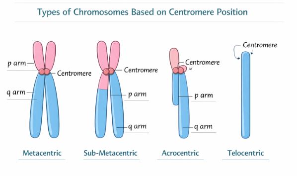
Every chromosome has a pinched-in “waist” called the primary constriction or Centromere. On the sides of the centromere are disc-shaped protein plates called kinetochores, where spindle fibers attach during division. Depending on exactly where this centromere is located, we classify chromosomes into four distinct types:
- Metacentric: Centromere is exactly in the middle. Creates two perfectly equal arms (looks like a ‘V’).
- Sub-metacentric: Centromere is slightly off-center. One arm is a bit shorter than the other (looks like an ‘L’).
- Acrocentric: Centromere is very close to one end. Creates one extremely short arm and one very long arm (looks like a ‘J’).
- Telocentric: Centromere is at the absolute terminal tip. Only one arm is visible (looks like an ‘I’).
Sometimes, a chromosome has a secondary constriction at a fixed spot, creating a tiny knob-like piece at the end called a satellite.
Real-Life Examples to Understand the Cell
- The City Analogy: The easiest way to learn cell organelles is to imagine the cell as a fortified city. The Plasma Membrane is the city wall with security gates. The Nucleus is the City Hall or Mayor’s office where all blueprints (DNA) are kept. The Mitochondria are the power plants generating electricity (ATP). The Endoplasmic Reticulum is the industrial highway network making goods. The Golgi apparatus is the Amazon fulfillment center, packaging and shipping those goods. The Lysosomes are the waste management and recycling crew keeping the city clean.
- Antibiotic Resistance: You hear doctors saying we shouldn’t overuse antibiotics. Why? Because bacteria have those tiny extra DNA rings called Plasmids. If a bacterium survives a dose of antibiotics, it encodes the “resistance recipe” into its plasmid. It can then pass this plasmid to its neighbors, creating a superbug!
Key Takeaways & Summary
- The cell is the fundamental structural and functional unit of life. Schleiden, Schwann, and Virchow gave us the complete Cell Theory.
- Cells are broadly divided into Prokaryotes (no true nucleus, no membrane-bound organelles) and Eukaryotes (distinct nucleus, highly compartmentalized).
- The cell membrane is a fluid, dynamic structure described by the Fluid Mosaic Model, consisting of a lipid bilayer with embedded proteins.
- The Endomembrane system includes ER, Golgi, lysosomes, and vacuoles, which work in a coordinated assembly line.
- Mitochondria and chloroplasts are double-membrane organelles, have their own circular DNA, 70S ribosomes, and are the energy centers of the cell.
- Ribosomes are non-membrane bound protein factories. Eukaryotes have 80S, prokaryotes have 70S.
- The nucleus houses the genetic material in the form of chromatin, which condenses into structurally defined chromosomes during cell division.
Common Student Misconceptions
Misconception 1: Prokaryotes do not have DNA.
Correction: A very dangerous mistake for your exams! Prokaryotes absolutely have DNA. It is essential for life. Their DNA is just not enclosed inside a protective nuclear membrane; it floats freely in a central region called the nucleoid.
Misconception 2: Plant cells have chloroplasts, so they don’t need mitochondria.
Correction: Incorrect! Chloroplasts make the food (glucose) using sunlight. But to actually extract usable energy (ATP) from that food to stay alive, especially at night or in the roots, plant cells desperately need mitochondria, just like animal cells do.
Misconception 3: The cell wall and cell membrane are the same thing.
Correction: Not at all. The cell membrane is living, flexible, and selectively permeable—all cells have it. The cell wall is a dead, rigid outer layer found only outside the membrane in plants, fungi, and bacteria for structural support.
Practice Set: Test Your Knowledge (CBSE Pattern)
Very Short Answer Questions (1 Mark)
Q1. Who coined the phrase “Omnis cellula-e cellula” and what does it mean?
Answer: Rudolf Virchow coined this phrase. It translates to “all cells arise from pre-existing cells,” which completed the cell theory.
Q2. Why is the tail region of the lipid bilayer inside the plasma membrane?
Answer: The lipid tails are hydrophobic (water-hating) nonpolar hydrocarbons. They face inward to remain protected from the watery (aqueous) environments inside and outside the cell.
Q3. What is a mesosome?
Answer: A mesosome is a specialized membranous structure found in prokaryotes, formed by the inward folding of the plasma membrane. It helps in respiration, secretion, and cell wall formation.
Short Answer Questions (2-3 Marks)
Q4. Mitochondria and chloroplasts are not considered part of the endomembrane system. Why?
Answer: The endomembrane system consists of organelles (ER, Golgi, lysosomes, vacuoles) whose functions are highly coordinated and linked together in a specific sequence. The functions of mitochondria (ATP production) and chloroplasts (photosynthesis) operate independently and are not coordinated with these components, hence they are excluded from the system.
Q5. Differentiate between Rough ER and Smooth ER based on structure and function.
Answer:
Structure: Rough ER has ribosomes attached to its outer surface, giving it a bumpy appearance. Smooth ER lacks ribosomes on its surface, appearing smooth.
Function: Rough ER is actively involved in the synthesis and secretion of proteins. Smooth ER is the major site for the synthesis of lipids and steroidal hormones.
Q6. How do neutral solutes move across the plasma membrane? Can polar molecules move the same way?
Answer: Neutral solutes move across the plasma membrane by simple diffusion along their concentration gradient (from higher to lower concentration). Polar molecules cannot move through the nonpolar lipid bilayer in the same way. They require specialized carrier proteins embedded in the membrane to facilitate their transport.
Long Answer Questions (5 Marks)
Q7. Describe the Fluid Mosaic Model of the plasma membrane with a clear explanation of its fluid nature. Mention one passive and one active transport mechanism.
Answer: The Fluid Mosaic Model was proposed by Singer and Nicolson in 1972. According to this model, the cell membrane consists of a continuous, quasi-fluid double layer of phospholipids. The polar heads face outwards, and the hydrophobic tails face inwards. Embedded within this lipid bilayer are proteins arranged in a mosaic pattern. Some proteins lie on the surface (peripheral), while others are deeply buried (integral).
Fluid Nature: The membrane is not solid. The quasi-fluid nature of the lipid allows proteins to move laterally within the overall bilayer. This fluidity is essential for functions like cell growth, endocytosis, and division.
Transport:
– Passive Transport: Movement of water across the membrane by diffusion without energy expenditure is called osmosis.
– Active Transport: Movement of ions against their concentration gradient (from low to high) using energy (ATP). Example: The Sodium-Potassium pump.
Q8. What is a centromere? How are chromosomes classified based on the position of the centromere? Explain in detail.
Answer: A centromere is a primary constriction present on every visible chromosome during cell division. It holds the two chromatids together and has disc-shaped structures called kinetochores on its sides for spindle attachment. Based on the position of the centromere, chromosomes are classified into four types:
1. Metacentric: The centromere is exactly in the middle of the chromosome, forming two equal arms.
2. Sub-metacentric: The centromere is located slightly away from the center, resulting in one shorter arm and one longer arm.
3. Acrocentric: The centromere is situated very close to one end of the chromosome, forming one extremely short arm and one very long arm.
4. Telocentric: The centromere is located at the very terminal end, appearing as if the chromosome has only one arm.
Case-Based / Competency-Based Question (4 Marks)
Q9. Read the situation and answer the questions.
A young scientist in a lab is given a tissue sample. Upon observing it under an electron microscope, she notes the presence of a distinct cell wall, a large central empty-looking sac taking up almost 80% of the cell volume, and numerous double-membrane organelles containing stacked thylakoids. However, she cannot find any centrioles.
(a) Identify whether this is a plant cell or an animal cell. Give two reasons from the text.
(b) Name the large central sac and the membrane enclosing it.
(c) What is the function of the organelles containing stacked thylakoids?
Answer:
(a) It is a plant cell. Reasons: Presence of a cell wall and plastids (indicated by thylakoids), and the absence of centrioles, which are typical characteristics of plant cells.
(b) The sac is the large central vacuole. The single membrane enclosing it is called the tonoplast.
(c) The organelles are chloroplasts. They contain chlorophyll pigments in their thylakoids, which trap light energy essential for the process of photosynthesis.
Assertion-Reason Question
Q10. For the following question, two statements are given—one labeled Assertion (A) and the other labeled Reason (R). Select the correct answer from the codes (a), (b), (c), and (d) as given below.
(a) Both A and R are true, and R is the correct explanation of A.
(b) Both A and R are true, but R is not the correct explanation of A.
(c) A is true, but R is false.
(d) A is false, but R is true.
Assertion (A): Lysosomes are commonly referred to as the suicidal bags of the cell.
Reason (R): Lysosomes contain highly active hydrolytic enzymes capable of digesting carbohydrates, proteins, lipids, and nucleic acids at an acidic pH.
Answer: (a). The assertion is true, and the reason is the exact correct explanation. Because they contain these powerful, destructive hydrolytic enzymes, if the lysosome bursts, it will digest its own cell components, earning the name “suicide bag.”
End of Notes.
Students, mastering the structures and functions of the organelles will build a solid foundation for everything else we study in biology. Draw the diagrams of the plant cell, animal cell, and mitochondria in your notebooks for practice!
Read Also:
Chapter 7- Structural Organisation in Animals
For official syllabus and textbooks, visit the
NCERT Official Website.