
Introduction | Setting the Stage for Biological Classification
Hello students! Welcome back to our journey through the living world. In our previous discussions, we learned how to name and group individual organisms. But today, we are going to look at the grand scheme of things. How do we organize the entire spectrum of life on Earth?
Imagine you have a massive library with millions of books dumped in a giant pile. Finding a specific science fiction novel would be impossible. You would naturally start sorting them: fiction versus non-fiction, then by genre, then by author. Since the dawn of human civilization, we have been trying to do the exact same thing with living organisms. Early humans classified plants and animals based on basic survival needs—what could be eaten, what provided shelter, and what could be used for clothing.
However, as science progressed, we needed a more logical, universally accepted system. In this chapter, we will explore the fascinating evolution of biological classification, moving from simple physical observations to complex cellular and genetic analysis. We will dive deep into the Five Kingdom Classification system, which completely revolutionized how we view life on our planet. Grab your notebooks, and let’s explore how biologists brought order to the beautiful chaos of nature!
1. The History and Evolution of Classification Systems
1.1 Aristotle’s Early Attempt
The very first scientific attempt to organize life was made by the ancient Greek philosopher Aristotle. He didn’t have microscopes or DNA sequencing, so he relied purely on simple, observable physical characteristics.
- Plants: He looked at their stems and sizes, dividing them simply into trees, shrubs, and herbs.
- Animals: His approach was even simpler here. He grouped animals based on whether they had red blood or did not have red blood.
While Aristotle’s system was a great starting point, it was incredibly limited. Think about it—grouping a bird and a snake together just because they both have red blood ignores massive differences in their anatomy and life cycles!
1.2 Linnaeus and the Two Kingdom System
Fast forward to the time of Carolus Linnaeus. He introduced a slightly more robust system, known as the Two Kingdom Classification, which simply divided all life into Kingdom Plantae (plants) and Kingdom Animalia (animals).
This system was easy to use and understand for a long time, but as our scientific tools improved, major cracks began to show. The Linnaean system failed miserably at addressing several crucial biological distinctions:
- It grouped single-celled (unicellular) and multi-celled (multicellular) organisms together.
- It placed organisms with a distinct nucleus (eukaryotes) alongside microscopic entities lacking a true nucleus (prokaryotes).
- It completely ignored the mode of nutrition, placing photosynthesizing green algae in the same group as non-photosynthesizing fungi.
Because countless organisms—like certain microbes or fungi—didn’t fit neatly into either the plant or animal box, scientists realized this two-kingdom model was highly inadequate. They needed a system that looked beyond just the “gross morphology” (outer appearance) and considered cellular anatomy, cell wall composition, how the organism feeds, and its evolutionary history.
1.3 R.H. Whittaker’s Five Kingdom Classification
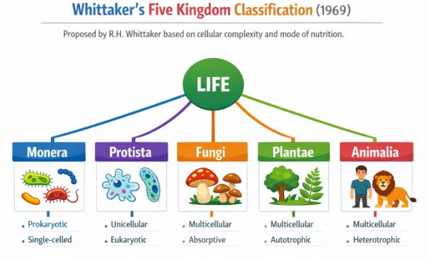
Figure-1: The Five Kingdom Classification system proposed by R.H. Whittaker in 1969, separating organisms based on cellular complexity and nutrition.
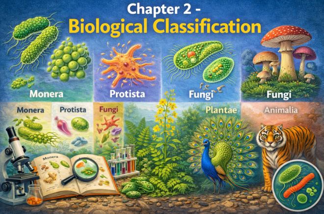
In the year 1969, an ecologist named R.H. Whittaker proposed a brilliant solution: the Five Kingdom Classification. This is the primary system we still study today. He created five distinct kingdoms: Monera, Protista, Fungi, Plantae, and Animalia.
Teacher’s Tip: If you get a question in the exam asking about the criteria Whittaker used, always list these five points:
- Cell Structure: Is it prokaryotic or eukaryotic?
- Body Organisation: Is it single-celled, a loose tissue, or a complex organ system?
- Mode of Nutrition: Does it make its own food (autotrophic) or rely on others (heterotrophic)?
- Reproduction: How does it multiply?
- Phylogenetic Relationships: How did it evolve over time?
This new system solved the old problems. For instance, bacteria and blue-green algae (which are prokaryotes) were finally separated from eukaryotic plants. Fungi, which have cell walls made of chitin and are heterotrophic, were rightfully removed from the plant kingdom (which have cellulosic walls and are autotrophic) and given their own exclusive kingdom.
2. Kingdom Monera: The World of Bacteria
Welcome to the invisible world! Kingdom Monera is completely composed of bacteria. These microscopic survivors are the most abundant living organisms on Earth, found practically everywhere—from a handful of garden soil to the deepest, darkest ocean trenches.
2.1 Shape and Structure
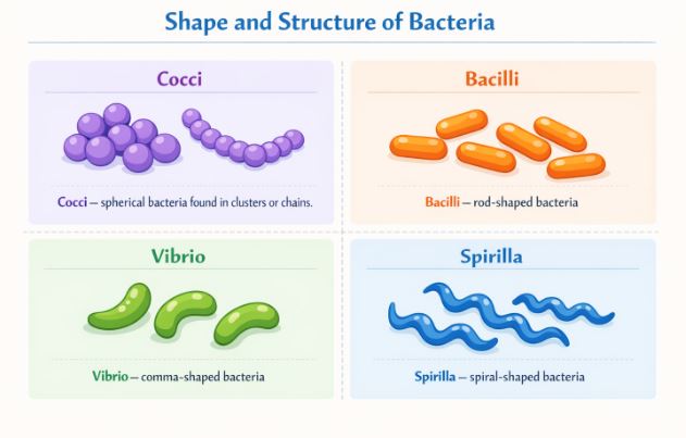
Figure-2: Bacteria are primarily grouped into four structural categories based on their physical shape.
Though bacteria look incredibly simple under a microscope, their internal chemistry and behavior are incredibly complex. They display the most extensive metabolic diversity of any group of organisms. Physically, we group them into four main shapes:
- Coccus (pl. cocci): Spherical or round-shaped.
- Bacillus (pl. bacilli): Rod-shaped, resembling small capsules.
- Vibrium (pl. vibrio): Comma-shaped.
- Spirillum (pl. spirilla): Spiral-shaped, like a corkscrew.
2.2 Archaebacteria (The Extremophiles)
Archaebacteria are the thrill-seekers of the microscopic world. They possess a highly unique cell wall structure that allows them to thrive in environments that would instantly kill other life forms.
- Halophiles: Love extreme salty environments.
- Thermoacidophiles: Thrive in boiling hot springs and highly acidic areas.
- Methanogens: Live in marshy swamplands and inside the guts of ruminant animals (like cows and buffaloes). Fun fact: When you see biogas being produced from cow dung, it’s actually these methanogens hard at work producing methane gas!
2.3 Eubacteria (The True Bacteria)
Eubacteria, or “true bacteria,” are characterized by their rigid cell walls and, if they can move, a whip-like structure called a flagellum.
Autotrophic Eubacteria: Some make their own food. Cyanobacteria (often called blue-green algae) have chlorophyll similar to green plants and perform photosynthesis. They often form colonies covered by a gelatinous sheath and can cause “blooms” in polluted waters. Some, like Nostoc and Anabaena, have special cells called heterocysts that can pull nitrogen right out of the air and fix it into the soil. Other autotrophs are chemosynthetic—they don’t use sunlight; instead, they oxidize inorganic chemicals like ammonia and nitrates to generate energy (ATP), playing a massive role in recycling nutrients.
Heterotrophic Eubacteria: These rely on organic matter and are the most abundant in nature. Many act as crucial decomposers, breaking down dead matter. They also impact our daily lives immensely: they help curdle milk into yogurt, produce vital antibiotics, and unfortunately, cause severe diseases like cholera, typhoid, and tetanus.
Reproduction: Bacteria generally multiply rapidly through a simple cell division process called fission. If conditions get tough (no food, extreme heat), they protect their DNA by forming tough spores.
Note on Mycoplasma: These are strange, tiny organisms under Monera that completely lack a cell wall. They hold the record for being the smallest known living cells and can surprisingly survive entirely without oxygen.
3. Kingdom Protista: The Microscopic Eukaryotes
If a single-celled organism has a true, well-defined nucleus and membrane-bound organelles (making it a eukaryote), it belongs in Kingdom Protista. Most protists live in aquatic environments. Interestingly, the boundaries of this kingdom are very blurry; what one scientist calls a photosynthetic protist, another might argue is a simple plant. Protista essentially acts as a massive evolutionary bridge connecting bacteria to the higher kingdoms of fungi, plants, and animals. Let’s examine the five major groups within this kingdom.
3.1 Chrysophytes (Diatoms and Golden Algae)
Found drifting passively in both fresh and marine water currents as plankton, these microscopic organisms are the ultimate glass-makers of nature. Diatoms have unique cell walls made of two thin, overlapping shells that fit together perfectly, much like a soapbox. Because these walls are embedded with tough silica, they are virtually indestructible. When diatoms die, their silica shells sink and accumulate on the ocean floor over billions of years, creating a gritty soil known as ‘diatomaceous earth’. We humans harvest this gritty soil for polishing and filtering oils and syrups. More importantly, diatoms perform so much photosynthesis that they are considered the chief ‘producers’ in the oceans.
3.2 Dinoflagellates
These are mostly marine organisms that look like tiny armored tanks because their cell walls have stiff cellulose plates on the outside. They possess two flagella—one running lengthwise and the other horizontally in a groove between the plates. Depending on their internal pigments, they can appear yellow, green, brown, blue, or red.
Teacher’s Warning: Some red dinoflagellates, like Gonyaulax, can multiply so explosively that they turn the sea completely red—a phenomenon known as a “red tide”. During a red tide, they release massive amounts of highly potent toxins that can kill huge populations of fish and other marine life.
3.3 Euglenoids
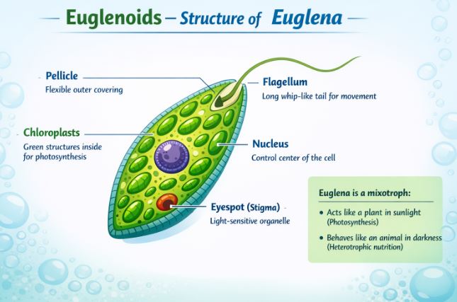
Found mostly in stagnant freshwater, euglenoids like Euglena are the rule-breakers of nature. They do not have a hard cell wall; instead, they are covered by a protein-rich, flexible layer called a pellicle. They have two flagella (one short, one long) for swimming. The coolest thing about them? They are mixotrophic! When sunlight is available, their plant-like pigments perform photosynthesis. But throw them into the dark, and they suddenly act like tiny animals, hunting and eating other smaller microbes.
3.4 Slime Moulds
These are saprophytic (decay-eating) protists. They creep over rotting twigs and dead leaves, consuming organic debris. When the environment is favorable, they clump together into a massive, spreading structure called a plasmodium. If the environment turns harsh and dry, this plasmodium forms fruiting bodies that release highly resistant spores into the wind, allowing them to survive for years until conditions improve.
3.5 Protozoans (The “First Animals”)
Protozoans are entirely heterotrophic, living as predators or parasites, and are considered the primitive relatives of actual animals.
- Amoeboid Protozoans: Move and capture prey by extending false feet called pseudopodia (e.g., Amoeba). Some, like Entamoeba, live inside us as harmful parasites.
- Flagellated Protozoans: Use flagella to move. Parasitic types cause dangerous human diseases, such as Sleeping Sickness (caused by Trypanosoma).
- Ciliated Protozoans: Covered in thousands of tiny, beating hairs called cilia. They have a cavity called a gullet, and the coordinated beating of the cilia steers food-laden water right into their “mouth” (e.g., Paramoecium).
- Sporozoans: These form infectious spores during their life cycle. The most infamous is Plasmodium, the deadly parasite responsible for causing malaria in humans.
4. Kingdom Fungi: The Great Decomposers
Have you ever noticed fuzzy white or green patches on old bread, or seen a mushroom sprouting from the soil? You are looking at Kingdom Fungi. Fungi are a massive group of heterotrophic organisms that prefer warm, humid places to grow. This is exactly why we invented refrigerators—to keep our food cold and prevent fungal and bacterial spores from germinating!
4.1 Fungal Structure and Nutrition
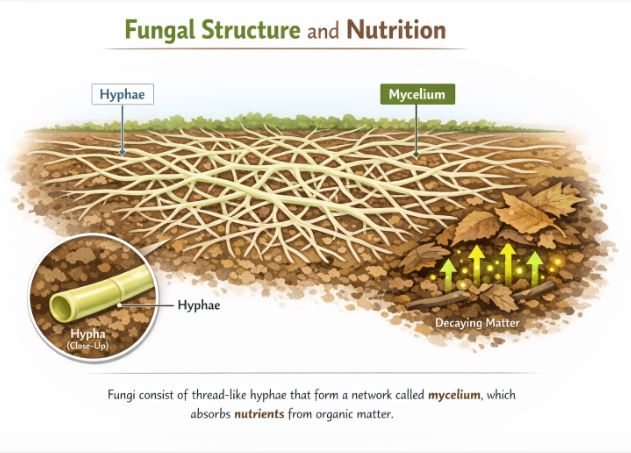
Except for yeast (which is unicellular and used to make bread and beer), all fungi are multicellular and have bodies made of long, thin, thread-like tubes called hyphae. A dense, tangled network of these hyphae is called a mycelium.
Some hyphae are just continuous tubes filled with cytoplasm and multiple nuclei (coenocytic), while others have cross-walls dividing them (septate). Their cell walls are unique because they are made of a tough substance called chitin along with polysaccharides.
Fungi cannot make food. Most act as saprophytes, absorbing soluble nutrients from dead, rotting matter. Some act as parasites, sucking nutrients from living plants and animals (like the Puccinia fungus that causes wheat rust). Others are team players, living as symbionts: when they partner with algae, we call it a lichen; when they partner with the roots of big trees, we call it mycorrhiza.
4.2 The Fungal Life Cycle and Reproduction
Fungi can reproduce vegetatively (by breaking apart or budding) and asexually via spores (like conidia or zoospores). But their sexual reproduction is quite dramatic and follows three distinct steps:
- Plasmogamy: The protoplasm (cell contents) of two mating cells fuse together.
- Karyogamy: The two distinct nuclei finally fuse to form a diploid cell.
- Meiosis: The diploid zygote undergoes reduction division to produce new, haploid spores inside special fruiting bodies.
Special Note: In certain higher fungi (Ascomycetes and Basidiomycetes), plasmogamy happens, but karyogamy is delayed. This creates an intervening stage where a single cell has two separate nuclei (n + n). This fascinating state is called a dikaryon, and the phase is the dikaryophase. Eventually, the nuclei fuse.
4.3 The Four Classes of Fungi
Biologists divide Kingdom Fungi into four main classes based on how their mycelium looks and how they produce sexual spores:
- Phycomycetes: Found in damp areas or as plant parasites. Their mycelium lacks cross-walls (aseptate). Example: Rhizopus (bread mould) and Albugo (mustard parasite).
- Ascomycetes (Sac Fungi): Can be unicellular (yeast) or multicellular (Penicillium). They produce sexual spores called ascospores inside tiny sacs (asci). Some, like truffles and morels, are highly prized edible delicacies.
- Basidiomycetes (Club Fungi): This class includes the classic mushrooms, puffballs, and bracket fungi. Their sexual spores (basidiospores) are produced externally on a club-shaped structure called a basidium. Examples: Agaricus (mushroom) and Ustilago (smut).
- Deuteromycetes (Imperfect Fungi): These are called “imperfect” because scientists have only ever observed their asexual reproduction phase. If we ever discover their sexual stage, they are immediately re-classified into Ascomycetes or Basidiomycetes. They reproduce using asexual spores called conidia. Example: Trichoderma.
5. A Brief Look at Plantae and Animalia
We will dive deeply into these kingdoms in the next chapters, but let’s summarize them:
Kingdom Plantae: Contains all eukaryotic, chlorophyll-bearing, autotrophic organisms. Their cell walls are primarily made of cellulose. While mostly autotrophic, a few rebel plants exist—like the Venus flytrap (partially heterotrophic/insect-eating) and Cuscuta (a pure parasite). A key feature of plants is their dual life cycle: they alternate between a diploid sporophyte phase and a haploid gametophyte phase, a phenomenon known as the alternation of generations.
Kingdom Animalia: Contains all multicellular, eukaryotic organisms that completely lack cell walls. Animals are purely heterotrophic; they must consume other organisms for food and digest it inside an internal cavity. They store their excess energy as glycogen or fat. Animals exhibit a holozoic mode of nutrition (ingesting solid food) and mostly possess complex sensory and nervous systems allowing for active locomotion.
6. The Exceptions: Viruses, Viroids, Prions, and Lichens
If you carefully read Whittaker’s Five Kingdom chart, you will notice some famous names are completely missing. Where do the viruses that cause cold and flu fit in? The answer: nowhere.
6.1 Viruses: The Borderline of Life
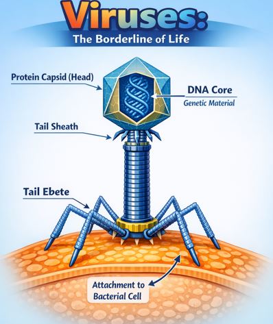
Viruses did not get a kingdom because they are not truly “living” cellular organisms. Outside a host body, a virus is just an inert, crystalline chemical structure. However, the moment they penetrate a living host cell, they hijack the host’s internal machinery to replicate themselves, usually destroying the host cell in the process.
A virus is fundamentally a nucleoprotein—it consists of a protein coat (called a capsid) enveloping a core of genetic material. Crucial rule: The genetic material can be RNA or DNA, but never both at the same time. Viruses are responsible for terrible diseases, from the common cold and influenza to smallpox and AIDS. Viruses that specifically hunt and infect bacteria are known as bacteriophages.
6.2 Viroids and Prions
Think a virus is small? Meet the Viroid. Discovered by T.O. Diener in 1971, viroids are even smaller infectious agents consisting only of free, low-molecular-weight RNA. They completely lack the protective protein coat found in viruses. They are famous for causing the potato spindle tuber disease.
Then we have Prions. These don’t even have DNA or RNA! Prions are simply abnormally folded proteins that can transmit severe neurological diseases. They are the culprits behind Bovine Spongiform Encephalopathy (BSE)—commonly known as “Mad Cow Disease” in cattle—and its human equivalent, CJD.
6.3 Lichens: Nature’s Perfect Marriage
A lichen is not a single organism. It is an intimate, mutually beneficial symbiotic association between an alga (the phycobiont) and a fungus (the mycobiont). The alga, being autotrophic, cooks up food via photosynthesis for both of them. In return, the tough fungus provides physical shelter and absorbs essential water and minerals for the alga. Their bond is so seamless that if you spot a lichen growing on a tree trunk, you would assume it is just one single organism.
Ecological importance: Lichens are incredibly sensitive to sulfur dioxide pollution. They absolutely refuse to grow in polluted areas, making them excellent natural indicators of air quality.
Real-Life Examples to Understand Concepts
- Diatomaceous Earth at Home: The same microscopic diatom shells we learned about in Kingdom Protista are actually used in some natural toothpastes as a mild abrasive to scrub away plaque, and in swimming pool filters to trap dirt!
- Fungal Antibiotics: When you get sick and a doctor prescribes Penicillin, you are using a chemical weapon originally created by an Ascomycete fungus (Penicillium) to defend its territory against invading bacteria.
- Biogas Plants in Villages: The biogas used for cooking in rural areas is completely dependent on Archaebacteria (methanogens). These bacteria break down the cow dung in anaerobic digesters and release methane gas as a byproduct.
Key Takeaways & Summary
- Biological classification evolved from simple morphological grouping (Aristotle) to the robust Five Kingdom system (Whittaker).
- Monera: Comprises all prokaryotic bacteria. They exhibit immense metabolic diversity and can be autotrophic or heterotrophic.
- Protista: Contains single-celled eukaryotes (Diatoms, Dinoflagellates, Euglenoids, Slime moulds, Protozoans). It forms an evolutionary link to higher kingdoms.
- Fungi: Multicellular, heterotrophic saprophytes or parasites with chitinous cell walls. Divided into Phycomycetes, Ascomycetes, Basidiomycetes, and Deuteromycetes.
- Plantae & Animalia: Eukaryotic autotrophs with cellulosic walls (plants) and multicellular, wall-less heterotrophs (animals).
- Viruses, Viroids, and Prions are non-cellular infectious agents that don’t fit into Whittaker’s cellular classification.
Common Student Misconceptions
Misconception 1: Blue-green algae belong to Kingdom Plantae or Protista.
Correction: Despite the word “algae” in their common name, blue-green algae (Cyanobacteria) are prokaryotic organisms. Because they lack a true nucleus, they strictly belong to Kingdom Monera.
Misconception 2: Viruses are considered the simplest living cells.
Correction: Viruses are not cells at all! They have no cellular machinery, cytoplasm, or metabolism of their own. They are considered non-cellular, existing on the boundary line between living and non-living things.
Practice Set: Test Your Knowledge (CBSE Pattern)
Very Short Answer Questions (1 Mark)
Q1. What is the main structural difference that allows Archaebacteria to survive extreme conditions?
Answer: Archaebacteria possess a fundamentally different cell wall structure compared to normal eubacteria, which grants them high resistance against extreme heat, salt, and acidity.
Q2. Why are Deuteromycetes commonly referred to as ‘imperfect fungi’?
Answer: They are called imperfect fungi because biologists have only discovered and observed their asexual or vegetative reproductive phases. Their sexual life cycle remains unknown.
Short Answer Questions (2-3 Marks)
Q3. Explain the phenomenon of ‘red tides’. Which organism is responsible?
Answer: ‘Red tides’ occur when certain red dinoflagellates (like Gonyaulax), which belong to Kingdom Protista, multiply rapidly in the ocean. Their immense numbers make the water appear red. This event is highly dangerous because the dinoflagellates release potent toxins that can kill huge quantities of marine life, including fishes.
Q4. How do viroids differ structurally from true viruses? Name a disease caused by viroids.
Answer: True viruses consist of a core of genetic material (DNA or RNA) enclosed inside a protective protein coat (capsid). Viroids, on the other hand, are composed purely of free, low-molecular-weight RNA and completely lack a protein coat. A famous disease caused by viroids is the potato spindle tuber disease.
Long Answer Questions (5 Marks)
Q5. Critically analyze why R.H. Whittaker’s Five Kingdom Classification was a major improvement over Linnaeus’s Two Kingdom Classification. Explain the primary criteria Whittaker used.
Answer: Linnaeus’s Two Kingdom system (Plantae and Animalia) was fundamentally flawed because it lumped together highly diverse organisms purely based on superficial traits. It failed to distinguish between prokaryotes (bacteria) and eukaryotes, unicellular and multicellular forms, and autotrophs and heterotrophs (like placing photosynthetic plants alongside absorptive fungi).
Whittaker’s system solved this confusion by establishing distinct kingdoms based on cellular reality. His primary criteria for classification were:
1. Cell structure: Separating prokaryotes (into Monera) from eukaryotes.
2. Body organisation: Differentiating unicellular organisms (Protista) from complex multicellular organisms.
3. Mode of nutrition: Separating autotrophs (Plantae) from ingestive heterotrophs (Animalia) and absorptive heterotrophs (Fungi).
4. Reproduction methods.
5. Phylogenetic (evolutionary) relationships.
By applying these criteria, fungi received their own kingdom, and the microscopic world was accurately categorized into Monera and Protista.
Q6. Describe the defining features of Kingdom Fungi, focusing on their somatic structure and the three major steps involved in their sexual reproductive cycle.
Answer: Kingdom Fungi comprises heterotrophic, eukaryotic organisms that typically possess cell walls made of chitin. Except for unicellular yeast, their body (somatic structure) consists of a network of long, slender thread-like tubes called hyphae, and the entire mass of hyphae is termed a mycelium. They absorb soluble organic matter (saprophytic).
When fungi reproduce sexually, the cycle follows three precise steps:
1. Plasmogamy: The fusion of protoplasm between two compatible mating cells (motile or non-motile).
2. Karyogamy: The fusion of the two parent nuclei to form a single diploid nucleus.
3. Meiosis: The diploid cell undergoes reduction division within a fruiting body, generating new haploid spores that will eventually grow into a new mycelium.
Case-Based / Competency-Based Question (4 Marks)
Q7. Read the following situation and answer the questions.
During a field trip to an unpolluted forest, a biology student notices crusty, grayish-green patches growing flat against the bark of mature trees. The teacher explains that this is not a single plant, but an incredible symbiotic partnership that serves as nature’s air quality monitor. However, the student observes none of these patches on the trees near the heavily industrialized highway on their way back.
(a) Identify the organism described in the passage.
(b) Name the two biological components that make up this organism. What specific roles do they play for each other?
(c) Why was this organism entirely absent on the trees near the industrial highway?
Answer:
(a) The organism is a Lichen.
(b) The components are an alga (phycobiont) and a fungus (mycobiont). The autotrophic alga prepares food for both through photosynthesis. The heterotrophic fungus provides a physical shelter structure and absorbs water and essential minerals from the environment for the alga.
(c) Lichens are highly sensitive biological indicators of air pollution, specifically sulfur dioxide. They cannot survive or grow in polluted environments, which is why they are absent near the industrial highway.
Assertion-Reason Question
Q8. For the following question, two statements are given—one labeled Assertion (A) and the other labeled Reason (R). Select the correct answer from the codes (a), (b), (c), and (d) as given below.
(a) Both A and R are true, and R is the correct explanation of A.
(b) Both A and R are true, but R is not the correct explanation of A.
(c) A is true, but R is false.
(d) A is false, but R is true.
Assertion (A): Organisms like Euglena pose a major classification challenge because they exhibit both plant-like and animal-like characteristics.
Reason (R): Euglenoids possess a rigid cellulosic cell wall like plants but move actively using cilia like animals.
Answer: (c) A is true, but R is false. The assertion is true; Euglena acts autotrophic in sunlight but heterotrophic in the dark. However, the reason is entirely false. Euglenoids do NOT have a rigid cellulosic cell wall; they have a flexible protein-rich pellicle, and they use flagella, not cilia, for movement.
End of Notes.
Students, remember that biological classification is a living science. As we learn more about DNA and cellular structures, our systems will continue to evolve. Keep reviewing these kingdoms and their key differences!
Read Also:
Chapter 1- The Living World
For official syllabus and textbooks, visit the
NCERT Official Website.