
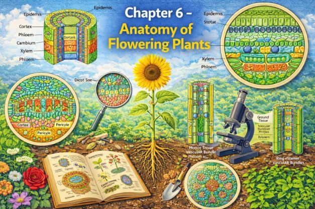
Introduction | Class 11 Biology Chapter 6 Anatomy of Flowering Plants notes
Hello students! Welcome back to our biology classroom. In our previous discussions, we explored the external features of plants—the shapes of leaves, the types of roots, and the beautiful arrangements of flowers. We learned that by simply looking at the external morphology of large living organisms, we can easily spot both structural similarities and striking variations. But today, we are going to do something different. Today, we are going to grab our metaphorical microscopes and look inside.
Think about building a house. From the outside, you see the paint, the windows, and the roof. But the true strength and function of that house depend on the internal wiring, the plumbing pipes, and the brick foundation hidden behind the walls. Plants are exactly the same! When we study the internal structure of a plant, we discover a whole new world of several similarities and fascinating differences. This scientific study of the internal structure of plants is known as anatomy.
Just like your body is made of cells that form tissues, which then form organs like your heart and lungs, plants follow a similar blueprint. In plants, cells act as the basic fundamental unit, which are grouped and organised into tissues, and these tissues further organise themselves into plant organs like roots, stems, and leaves. Interestingly, different organs within the very same plant will show remarkable differences in their internal structures. Furthermore, if we look at the group of flowering plants (angiosperms), we will notice that monocots and dicots are anatomically quite different from each other. These internal architectures aren’t random; they show brilliant adaptations designed to help the plant survive in diverse environments. Let’s dive into this hidden microscopic world!
1. The Tissue Systems
Before we look at the organs, we need to understand the building materials. Tissues in a plant body vary greatly depending on where they are located. This makes perfect sense, right? A tissue on the surface of a leaf has a very different job than a tissue buried deep inside a root. Therefore, their structure and their function are heavily dependent on their specific location.
Based on their structure and exactly where they are located, botanists categorize plant tissues into three primary tissue systems. These are:
- The Epidermal Tissue System: The protective outer skin.
- The Ground (or Fundamental) Tissue System: The main filling or packing material.
- The Vascular (or Conducting) Tissue System: The internal plumbing and transport system.
1.1 The Epidermal Tissue System
Imagine putting on a raincoat before stepping out into a storm. For a plant, the epidermal tissue system acts as that protective raincoat. It forms the extreme outer-most covering of the entire plant body. This system is comprised of three main components: the epidermal cells themselves, tiny pores called stomata, and various epidermal appendages like hairs and trichomes.
The epidermis acts as the outermost layer of the primary plant body. If you look at it under a microscope, you will see elongated, compactly arranged cells that form a continuous, unbroken layer. Usually, this protective wall is just a single layer thick. These epidermal cells are parenchymatous in nature; they contain a large central vacuole and have a small amount of cytoplasm lining their cell wall.
Now, to prevent the plant from drying out under the hot sun, the outside of the epidermis is frequently covered with a thick, waxy layer known as the cuticle, which is excellent at preventing water loss. Teacher’s Question: Would you expect to find a cuticle on a root? Think about it! Roots need to absorb water from the soil. If they were covered in wax, they would die of thirst! Therefore, the cuticle is entirely absent in roots.
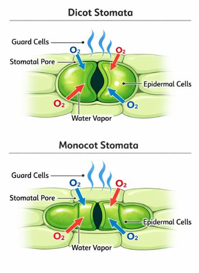
Figure-1: Notice the difference in the shape of guard cells. Bean-shaped are typical in dicots, while dumbbell-shaped are found in grasses.
Let’s talk about the breathing pores: Stomata. These are specialized structures located in the epidermis of leaves. They have a massive responsibility—they regulate the process of gaseous exchange (breathing in CO2, breathing out O2) and transpiration (water loss). Each individual stoma is guarded by two specialized cells known as guard cells, which enclose the stomatal pore.
Here is a fascinating anatomical difference: In most plants, guard cells are bean-shaped, but in grasses (which are monocots), the guard cells are distinctly dumb-bell shaped. The walls of these guard cells are unevenly thickened; the outer walls facing away from the pore are quite thin, while the inner walls facing towards the stomatal pore are highly thickened. Because these guard cells possess chloroplasts, they can perform photosynthesis and actively regulate the opening and closing of the stomata. Often, a few neighboring epidermal cells change their shape and size to help out the guard cells; we call these helper cells subsidiary cells. Together, the stomatal aperture, the guard cells, and the surrounding subsidiary cells make up what we call the stomatal apparatus.
Finally, the epidermis bears appendages. On roots, we find root hairs, which are simple, unicellular elongations of the epidermal cells designed to absorb water and minerals from the surrounding soil. On the stem, these epidermal hairs are called trichomes. Unlike root hairs, trichomes in the shoot system are usually multicellular. They can be soft or stiff, branched or unbranched, and sometimes even secretory. Their primary job? To help prevent water loss due to transpiration by trapping a layer of still air next to the plant surface.
1.2 The Ground Tissue System
If you take away the outer epidermis and the inner vascular pipelines, everything else that remains constitutes the ground tissue. It forms the main bulk of the plant. You can think of it as the fleshy part of an apple or the stuffing inside a pillow.
This system consists primarily of simple tissues—namely parenchyma, collenchyma, and sclerenchyma. In primary stems and roots, you will usually find parenchymatous cells occupying regions known as the cortex, pericycle, pith, and medullary rays. In the leaves, the ground tissue has a special name. Because it consists of thin-walled cells heavily packed with chloroplasts for photosynthesis, the ground tissue of the leaf is called the mesophyll.
1.3 The Vascular Tissue System
This is the plant’s transportation network. The vascular system consists of complex tissues—specifically the xylem (which transports water) and the phloem (which translocates food). Together, the xylem and phloem bundle up to constitute what we call vascular bundles.
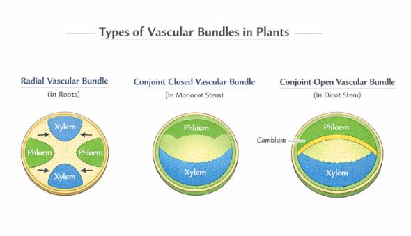
Figure-2: The arrangement of Xylem and Phloem determines the type of vascular bundle. Note the presence of Cambium in the open type.
The arrangement of these bundles varies. Let’s look at the key types:
- Radial Arrangement: When the xylem and phloem within a bundle are arranged in an alternate manner along different radii, it is called radial. This type of arrangement is a classic feature found in roots.
- Conjoint Arrangement: In this type, the xylem and phloem are jointly situated alongside each other on the exact same radius of the vascular bundle. This is the common setup you will find in stems and leaves. Usually, in conjoint bundles, the phloem is located exclusively on the outer side of the xylem.
Now, let’s talk about growth potential. In dicotyledonous stems, there is a special layer of actively dividing cells called cambium present right between the phloem and the xylem. Because they possess this cambium, these vascular bundles have the magical ability to form secondary xylem and secondary phloem tissues later in life (which creates wood!). Hence, they are referred to as open vascular bundles. On the flip side, in monocotyledons, the vascular bundles have no cambium present. Since they lack the machinery to form secondary tissues, they are simply referred to as closed vascular bundles.
2. Anatomy of Roots: Dicots vs. Monocots
To truly understand how plant organs are organized, botanists cut very thin slices across the mature zones of these organs. We call this a transverse section (T.S.). Let’s compare the transverse sections of dicot and monocot roots.
2.1 The Dicotyledonous Root (e.g., Sunflower Root)
Imagine traveling from the soil right to the center of a sunflower root. Here is the tissue organization you would encounter:
- Epiblema: This is the outermost protective layer. Many cells of the epiblema protrude outwards to form the delicate, unicellular root hairs.
- Cortex: Beneath the epiblema is a wide zone called the cortex. It consists of several layers of thin-walled parenchyma cells, complete with intercellular spaces to allow water to flow freely.
- Endodermis: This is the innermost layer of the cortex, acting like a biological border checkpoint. It is a single layer of barrel-shaped cells packed tightly without any intercellular spaces. The most critical feature here is that the radial and tangential walls of these endodermal cells have a heavy deposition of a waxy, water-impermeable material called suberin. This deposition takes the form of bands known as Casparian strips. This prevents water from leaking back out of the vascular system!
- Pericycle: Just inside the endodermis lies a few layers of thick-walled parenchymatous cells called the pericycle. This layer is very important because it is here that lateral roots and the vascular cambium are initiated during secondary growth.
- Vascular Bundles & Pith: In a dicot root, there are usually just two to four xylem and phloem patches. The central area, called the pith, is very small or sometimes completely inconspicuous. The parenchymatous cells sitting between the xylem and phloem are termed the conjuctive tissue. Later in life, a cambium ring develops between these patches.
Note: All the tissues situated on the inner side of the endodermis (pericycle, vascular bundles, and pith) collectively constitute what we call the stele.
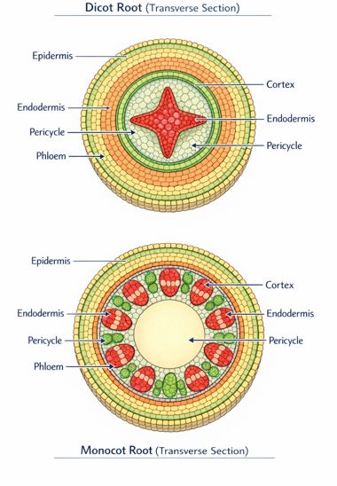
2.2 The Monocotyledonous Root
The anatomy of a monocot root shares many similarities with the dicot root. If you look at it, it also has an epidermis, cortex, endodermis, pericycle, vascular bundles, and a pith.
However, a smart student will spot the differences immediately. While a dicot root has very few xylem bundles (usually 2 to 4), a monocot root typically has more than six xylem bundles! We call this a polyarch condition. Furthermore, the pith right in the center is large and extremely well developed. Also, remember our rule about cambium? Monocotyledonous roots do not have it, which means they absolutely do not undergo any secondary growth. They stay relatively slender for their whole lives.
3. Anatomy of Stems: Dicots vs. Monocots
Now, let’s move up above the soil and cut a transverse section of young stems. The structural differences here are quite dramatic!
3.1 The Dicotyledonous Stem
If we look at a typical young dicot stem, the outermost protective layer is, of course, the epidermis. It is covered with a thin layer of cuticle and may bear stomata and trichomes. Just below this lies the cortex, but unlike the root, the stem’s cortex is complex and divided into three distinct sub-zones:
- Hypodermis: The outermost cortical zone, situated right below the epidermis. It consists of a few layers of collenchymatous cells that act like biological scaffolding, providing vital mechanical strength to the growing, young stem.
- Cortical Layers: Below the hypodermis, we find rounded, thin-walled parenchymatous cells with highly conspicuous intercellular spaces.
- Endodermis (Starch Sheath): The innermost layer of the cortex is the endodermis. In dicot stems, the cells of this layer are heavily packed with starch grains, which is why this layer is frequently referred to as the starch sheath.
Moving deeper, we find the pericycle sitting on the inner side of the endodermis, right above the phloem. It looks like semi-lunar (half-moon) patches made of tough sclerenchyma. Between the vascular bundles, there are radial layers of parenchymatous cells known as medullary rays.
The defining feature: If you look at a dicot stem under a microscope, you will see a large number of vascular bundles arranged in a beautiful, perfect ring. This ‘ring’ arrangement is the absolute hallmark characteristic of a dicot stem. Each bundle in this ring is conjoint, open (has cambium), and features endarch protoxylem. Finally, a massive central portion composed of rounded, parenchymatous cells with large spaces constitutes the pith.
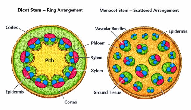
3.2 The Monocotyledonous Stem
Monocot stems do things very differently. Firstly, their hypodermis is not made of collenchyma; instead, they have a tough, sclerenchymatous hypodermis.
Secondly, you won’t find a neat ring of vascular bundles here. Instead, there are a large number of vascular bundles completely scattered throughout a large, conspicuous parenchymatous ground tissue. Every single one of these scattered vascular bundles is enclosed by its own sclerenchymatous bundle sheath.
These bundles are conjoint and closed (no cambium for secondary growth). An interesting observation is that the vascular bundles located towards the periphery (the edges) are generally much smaller than the ones located squarely in the center. Also, inside these bundles, phloem parenchyma is notably absent, and there are distinct water-containing cavities present.
4. Anatomy of Leaves: Dicots vs. Monocots
Leaves are the solar panels of the plant. Their internal structure is heavily optimized for capturing sunlight and managing gas exchange while minimizing water loss. Let’s examine vertical sections through the lamina of leaves.
4.1 The Dorsiventral (Dicotyledonous) Leaf
A dicot leaf is called “dorsiventral” because its upper (dorsal) side is structurally different from its lower (ventral) side. A vertical section reveals three main parts: the epidermis, the mesophyll, and the vascular system.
The epidermis covers both the upper surface (known as the adaxial epidermis) and the lower surface (the abaxial epidermis). Both have a conspicuous cuticle to block water loss. However, to prevent excessive evaporation under the direct sun, the lower abaxial epidermis generally bears far more stomata than the upper adaxial epidermis. In fact, the upper surface might completely lack stomata!.
The green tissue packed between the upper and lower epidermis is called the mesophyll. This is the photosynthesis factory of the leaf, made of parenchyma cells packed with chloroplasts. In dicot leaves, this mesophyll is highly differentiated into two distinct types of cells:
- Palisade Parenchyma: Placed adaxially (towards the upper surface). These cells are elongated and arranged vertically, completely parallel to one another like tightly packed columns. This helps them absorb maximum sunlight.
- Spongy Parenchyma: Situated right below the palisade cells, extending all the way to the lower epidermis. These cells are oval or round and very loosely arranged. Because they are loose, there are numerous large air cavities and spaces between them, allowing gases (CO2 and O2) to diffuse easily towards the stomata.
The vascular system can be seen inside the veins and the midrib. If you recall the outside of a dicot leaf, it has reticulate (net-like) venation. Because veins vary in thickness in reticulate venation, the sizes of the vascular bundles inside the leaf also vary greatly depending on the size of the vein. These bundles are securely surrounded by a thick-walled layer of cells called the bundle sheath.
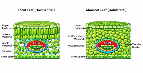
4.2 The Isobilateral (Monocotyledonous) Leaf
A monocot leaf (like grass or maize) is called “isobilateral” because both its surfaces are relatively similar and receive equal amounts of sunlight. Its anatomy shares many traits with the dicot leaf, but with a few very important characteristic differences.
Firstly, the distribution of breathing pores is equal. In an isobilateral leaf, stomata are present on both the upper and lower surfaces of the epidermis. Secondly, the internal solar panels are not split into specialized groups. The mesophyll is completely uniform and is not differentiated into palisade and spongy parenchyma.
Monocot leaves, specifically in grasses, have a brilliant trick for surviving droughts. Certain adaxial (upper) epidermal cells that lie along the veins modify themselves to become large, empty, colorless cells. These are known as bulliform cells. When rain falls and these cells absorb water, they become turgid (swollen), unrolling the leaf so its surface is fully exposed to light. However, when there is water stress and the plant is thirsty, these cells become flaccid (limp). This causes the entire leaf to curl inwards, hiding the stomata and minimizing water loss. Nature is genius!
Lastly, because monocot leaves feature parallel venation, their veins are mostly uniform in size. This is reflected internally, as you will see near-similar sizes of vascular bundles in vertical sections, except for the main central veins.
Real-Life Examples to Understand Anatomy
- The City Traffic System Analogy: Think of the plant’s tissue systems like a city. The Epidermal system is the city wall and gates (stomata) controlling who comes in and out. The Ground tissue represents the residential and industrial areas where actual work (like photosynthesis and food storage) happens. The Vascular system is the network of highways and pipelines. The Xylem is the water pipeline rushing water up from the roots, and the Phloem is the delivery truck network taking food from the leaf factories to the rest of the city.
- The Raincoat and the Sponge: The waxy cuticle on leaves and stems acts like a raincoat. But why don’t roots have it? Because roots act like a sponge. If you wrapped a sponge in a raincoat, it couldn’t soak up spills. Roots need to remain bare to absorb soil water efficiently.
Key Takeaways & Summary
- A plant is anatomically constructed from different kinds of tissues. These tissues handle primary functions like food assimilation, storage, transportation of water/minerals, and mechanical support.
- There are exactly three major tissue systems: epidermal, ground, and vascular.
- The epidermal system features epidermal cells, stomata, and appendages (hairs/trichomes).
- The ground tissue forms the massive bulk of the plant and is structurally divided into the cortex, pericycle, and pith.
- Vascular bundles (Xylem + Phloem) conduct water, minerals, and food. Their arrangement (radial vs conjoint) and the presence/absence of cambium define their type.
- Dicotyledonous and monocotyledonous plants display marked variations internally. They differ drastically in the type, location, and number of their vascular bundles.
- Only dicotyledonous roots and stems possess the ability to undergo secondary growth (making wood) due to the presence of cambium.
Common Student Misconceptions
Misconception 1: “All plant parts are covered by a thick waxy cuticle to prevent drying out.”
Correction: While it’s true for stems and leaves, roots absolutely do not have a cuticle! If a root had a waterproof waxy layer, it would be physically impossible for it to absorb water and minerals from the soil.
Misconception 2: “Monocot plants can grow into giant, thick wooden trees.”
Correction: Remember that monocot stems have “closed” vascular bundles, meaning they completely lack a cambium layer. Without cambium, they cannot produce secondary xylem (wood). This is why grasses, wheat, and maize stems stay relatively thin and green!
Practice Set: Test Your Knowledge (CBSE Pattern)
Very Short Answer Questions (1-2 Marks)
Q1. A student observes a transverse section of a stem under a microscope. They notice that the vascular bundles are conjoint, completely scattered throughout the ground tissue, and are surrounded by a sclerenchymatous bundle sheath. Furthermore, phloem parenchyma is absent. What type of stem has the student identified?
Answer: The student has identified a monocotyledonous stem. The scattered arrangement of vascular bundles and the absence of phloem parenchyma are hallmark features of a monocot stem.
Q2. How is the study of plant anatomy practically useful to us?
Answer: Studying plant anatomy helps us understand how plants adapt to different environments (like developing bulliform cells for drought). It allows us to distinguish between monocots and dicots, understand the quality of wood for timber, and identify plants in agriculture and forensics based on internal structures.
Short Answer Questions (3-4 Marks)
Q3. Explain the term ‘stomatal apparatus’. Explain the difference between the guard cells of a typical dicot and a grass plant.
Answer: The stomatal aperture, the two surrounding guard cells, and the adjacent specialized subsidiary cells together constitute the stomatal apparatus. In most dicot plants, the guard cells are distinctly bean-shaped. However, in grasses (which are monocots), the guard cells uniquely take on a dumb-bell shape.
Q4. Name the three basic tissue systems found in flowering plants. Provide the names of the specific tissues that fall under each of these systems.
Answer: The three basic tissue systems are:
1. Epidermal Tissue System: Includes epidermal cells, stomata, and appendages (trichomes and root hairs).
2. Ground Tissue System: Includes simple tissues like parenchyma, collenchyma, and sclerenchyma. (Forms the cortex, pericycle, pith, and leaf mesophyll).
3. Vascular Tissue System: Includes the complex conducting tissues, namely Xylem and Phloem.
Long Answer Questions (5 Marks)
Q5. Describe the internal structure of a dorsiventral (dicotyledonous) leaf in detail. Explain how its internal anatomy is suited for its function.
Answer: A dorsiventral leaf displays three main structural parts: the epidermis, the mesophyll, and the vascular system.
1. Epidermis: It covers both the upper (adaxial) and lower (abaxial) surfaces, coated with a protective cuticle. The lower surface typically bears significantly more stomata to allow gas exchange while being hidden from direct sun heat, minimizing water loss.
2. Mesophyll: This is the parenchymatous ground tissue responsible for photosynthesis. It is highly differentiated into two types:
a) Palisade parenchyma: Located adaxially, consisting of elongated, vertically parallel cells. Being near the top, they capture maximum sunlight.
b) Spongy parenchyma: Located below the palisade cells, consisting of loosely arranged oval cells with large air cavities. These spaces facilitate the easy diffusion of CO2 and O2 between the stomata and the photosynthesizing cells.
3. Vascular System: The vascular bundles are located in the veins and midrib, surrounded by thick-walled bundle sheath cells. Because dicots have reticulate venation, the vascular bundles vary greatly in size depending on the vein’s thickness. They efficiently supply water to the mesophyll and transport manufactured food away.
Q6. How would you ascertain whether a newly cut transverse section of a young stem from your school garden belongs to a monocot plant or a dicot plant? Give clear anatomical reasons to support your observation.
Answer: To determine if the stem is dicot or monocot, I would observe the arrangement and structure of the vascular bundles and the ground tissue under a microscope.
If it is a Dicot Stem:
– The vascular bundles will be arranged in a distinct, perfect ‘ring’.
– The bundles will be “open” (meaning a layer of cambium is present between xylem and phloem).
– The ground tissue will be clearly differentiated into cortex, endodermis, pericycle, and a large central pith.
– The hypodermis will be made of collenchyma cells.
If it is a Monocot Stem:
– The vascular bundles will be heavily scattered throughout the entire stem section.
– The bundles will be “closed” (absolutely no cambium present).
– The ground tissue will be a single, large, undifferentiated mass (no clear cortex or pith).
– The hypodermis will be made of sclerenchyma cells, and each vascular bundle will be enclosed by a sclerenchymatous bundle sheath.
Case-Based / Competency-Based Question (4 Marks)
Q7. Read the following situation and answer the questions.
A farmer noticed that during the intense, dry summer months, the leaves of his maize crop (a grass) began to roll inwards, taking a cylindrical shape. However, the leaves of his nearby sunflower crop remained flat, though slightly wilted. He asked his daughter, a biology student, to explain this phenomenon.
(a) Anatomically, what specific cells in the maize leaves are responsible for this rolling action?
(b) Where exactly are these specialized cells located on the leaf?
(c) Explain the mechanism of how these cells cause the leaf to curl during a dry summer.
(d) Why didn’t the sunflower leaves show this exact same inward rolling behavior?
Answer:
(a) The cells responsible for this action are called bulliform cells.
(b) They are found in monocots (grasses) and are modified adaxial (upper) epidermal cells situated alongside the veins.
(c) During dry summer months (water stress), the bulliform cells lose water and become completely flaccid (limp). This loss of turgor pressure causes the leaf surface to curl inwards, which safely hides the stomata and prevents further excessive water loss.
(d) The sunflower is a dicotyledonous plant. Its leaves are dorsiventral and do not possess these specialized bulliform cells in their upper epidermis, hence they cannot roll up in the same manner.
Assertion-Reason Question
Q8. For the following question, two statements are given—one labeled Assertion (A) and the other labeled Reason (R). Select the correct answer from the codes (a), (b), (c), and (d) as given below.
(a) Both A and R are true, and R is the correct explanation of A.
(b) Both A and R are true, but R is not the correct explanation of A.
(c) A is true, but R is false.
(d) A is false, but R is true.
Assertion (A): The inner boundary of the cortex in a dicot root acts as a water-tight checkpost, preventing water from leaking back out of the vascular tissue.
Reason (R): The endodermal cells of the dicot root possess Casparian strips, which are heavy depositions of water-impermeable, waxy suberin on their radial and tangential walls.
Answer: (a) Both A and R are true, and R is the correct explanation of A. The innermost layer of the cortex is the endodermis. It is the presence of suberin in the Casparian strips that makes this cellular layer impermeable to water, perfectly explaining why it acts as a watertight boundary for the stele.
End of Notes.
Students, make sure you practice drawing the transverse sections of the roots and stems. In anatomy, a neat diagram is worth a thousand words. Happy studying!
Read Also:
Chapter 5- Morphology of Flowering Plants
For official syllabus and textbooks, visit the
NCERT Official Website.