
Start Chapter MCQ Quiz
Comprehensive Guide: Structural Organisation in Animals (Frog) Class 11 Notes
Introduction | Class 11 Biology Chapter 7
Hello students! Welcome back to our biology classroom. In our previous discussions, we explored the massive variety of organisms that make up the animal kingdom, ranging from the simplest single-celled creatures to highly complex multicellular beings. Today, we are going to dive deep into how these living machines are built from the inside out.
Think about a tiny, unicellular organism for a moment. That one single cell is the ultimate multitasker. It handles digestion, respiration, and even reproduction all by itself. But what happens when organisms get larger and more complex? You cannot rely on just one cell to do all the heavy lifting. In complex multicellular animals, these basic life functions are divided among different groups of specialized cells, working together in a highly organized and synchronized manner.
Let’s look at a simple organism like the Hydra. Even though it’s relatively simple, its body is composed of different types of cells, and there can be thousands of cells belonging to each specific type. Now, scale that up to the human body! You are made up of billions of cells, all cooperating to perform the myriad functions that keep you alive, studying, and playing.
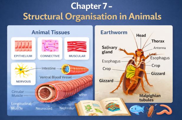
But how do all these billions of cells avoid chaos? They organize! As you might recall from earlier classes, in multicellular animals, a group of similar cells, along with the intercellular substances between them, team up to perform a specific function. We call this highly cooperative organization a tissue. You might find it fascinating that despite the incredible complexity of animals, all of them are constructed using just four basic types of tissues! When these basic tissues arrange themselves in specific proportions and patterns, they form an organ—like your stomach, your lungs, your heart, or your kidneys. Furthermore, when two or more organs collaborate chemically or physically to achieve a common goal, they form an organ system (like your digestive or respiratory system). This beautifully exhibits the “division of labour,” ensuring the whole body survives and thrives.
1. Organ and Organ System: The Evolutionary Trend
This beautiful hierarchy—where tissues build organs, and organs associate to form organ systems—is absolutely essential for coordinating the activities of the millions or billions of cells inside a multicellular organism. Every single organ in our body is constructed from one or more types of tissues. Take our heart, for example. It’s a fantastic pump that utilizes all four fundamental tissue types: epithelial, connective, muscular, and neural.
When scientists carefully study the complexity of organs and organ systems across different animals, they notice a distinct, observable pattern of improvement and adaptation over time. We refer to this pattern as an evolutionary trend (a concept you will explore deeply in Class XII).
Morphology vs. Anatomy
Before we jump into studying our model organism for this chapter, let’s clear up two important terms that students often mix up:
- Morphology: This is the study of form or externally visible features. If we are talking about plants or microbes, morphology simply means their external look. For animals, morphology refers to the external appearance of their body parts or organs.
- Anatomy: This term is conventionally used when we study the morphology of internal organs in animals. If you have to open the animal up to see it, you are studying anatomy!
In this chapter, we are going to master both the morphology and the anatomy of a very famous vertebrate: the frog. Let’s begin!
2. Meet the Frog (Rana tigrina)
Frogs are fascinating creatures that lead a double life. They are perfectly capable of living on land as well as in freshwater, which is why they belong to the class Amphibia under the phylum Chordata.
If you explore the ponds and fields in India, the most common species of frog you will encounter is Rana tigrina, commonly known as the Indian bullfrog.
2.1 Temperature Control and Survival Secrets
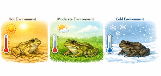
Unlike you and me, frogs do not have a built-in thermostat. They do not maintain a constant body temperature; instead, their internal temperature fluctuates based on the temperature of their surrounding environment. We call such animals cold-blooded or poikilotherms.
Have you ever tried to spot a frog in the grass and suddenly realized it blended in perfectly? Frogs have an incredible ability to change their color to hide from predators in grasses or on dry land. This clever trick of blending into the background is called camouflage , and this type of protective coloration is specifically known as mimicry.
Furthermore, you probably haven’t seen many frogs hopping around during the scorching peak of summer or the freezing dead of winter. To protect themselves from extreme heat and cold, they dig deep burrows and take shelter.
- The summer sleep to escape the heat is known as aestivation.
- The winter sleep to escape the cold is known as hibernation.
Think of it like putting your phone on “power saving mode” when conditions are tough!
3. Morphology: The External Features
Let’s look at the outside of the frog. If you have ever touched one, you know their skin feels smooth, wet, and highly slippery. This is due to the constant secretion of mucus. A frog must always maintain its skin in a moist condition. Here is a mind-blowing fact: Frogs never drink water! Instead, they absorb all the moisture they need directly through their skin.
Color-wise, the frog is generally olive green on its dorsal (back) side with dark, irregular spots. If you flip it over, the ventral (belly) side is a uniform, pale yellow.
3.1 Body Divisions and Features
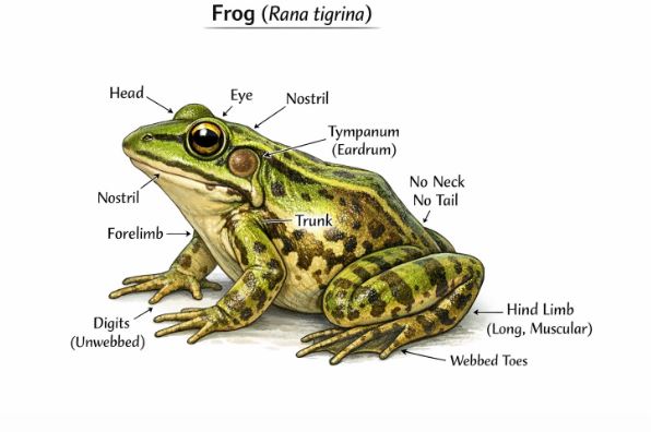
The body of an adult frog is very compact and is divided into just two main parts: the head and the trunk. They do not have a neck, nor do they have a tail.
- Head region: Just above the mouth, they have a pair of nostrils. Their eyes are quite distinctive—they are bulged outwards and are protected by a special transparent covering called a nictitating membrane, which acts like built-in swimming goggles to protect their eyes while underwater. Right behind the eyes on either side, you will find a membranous structure called the tympanum (the eardrum), which picks up sound signals.
- Limbs: The forelimbs and hind limbs are crucial tools for swimming, leaping, walking, and burrowing. The hind limbs are much larger and far more muscular than the forelimbs. Additionally, the hind limbs end in five digits and feature webbed feet to aid in swimming, whereas the smaller forelimbs end in only four digits.
3.2 Spotting the Difference: Sexual Dimorphism
If I place two frogs in front of you, how do you tell the boy from the girl? Frogs exhibit sexual dimorphism, meaning males and females have distinct physical differences. Male frogs are easily distinguished because they possess sound-producing vocal sacs (which they use to croak loudly during mating season) and a special copulatory pad located on the first digit of their forelimbs. Both of these features are completely absent in female frogs.
4. Anatomy: Exploring the Internal Systems
Now, let’s step into the fascinating world of the frog’s internal anatomy. The body cavity of a frog accommodates several well-developed organ systems, including the digestive, respiratory, circulatory, nervous, excretory, and reproductive systems. We will explore them one by one.
4.1 The Digestive System
The frog’s digestive system consists of the alimentary canal and associated digestive glands. Here is a very important conceptual question: Why is a frog’s alimentary canal relatively short? Because frogs are strict carnivores (meat-eaters). Meat is easier to break down than plant matter, so the length of their intestine is naturally reduced.
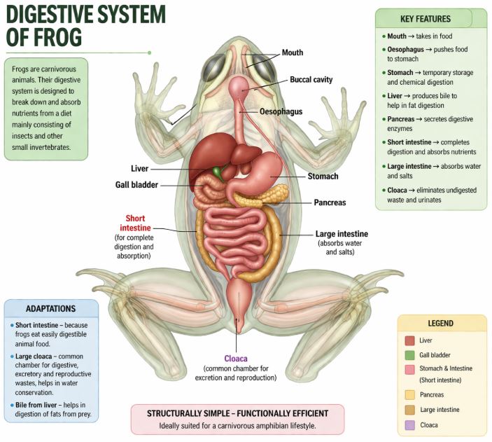
The Journey of Food:
- The frog captures its insect prey using its specialized bilobed tongue.
- The mouth opens into the buccal cavity, leading through the pharynx into a short tube called the oesophagus.
- The oesophagus opens into the stomach. Here, digestion heavily relies on Hydrochloric Acid (HCl) and gastric juices secreted by the stomach walls.
- This partially digested, soupy food mixture is called chyme. It passes from the stomach into the duodenum, the first section of the small intestine.
- In the duodenum, two important digestive juices arrive via a common bile duct: Bile (secreted by the liver and stored in the gall bladder) and pancreatic juices (produced by the pancreas). Bile works to emulsify fats, while pancreatic juices break down carbohydrates and proteins.
- Final digestion occurs in the intestine. The inner walls of the intestine feature numerous tiny finger-like folds known as villi and microvilli, which massively increase the surface area to absorb the digested nutrients.
- Finally, any undigested solid waste moves into the rectum and is expelled to the outside through a common exit chamber called the cloaca.
4.2 The Respiratory System
Frogs are amphibians, so naturally, they have dual mechanisms for breathing! They respire differently on land and in water.
- In Water (Cutaneous Respiration): When submerged, the frog uses its skin as an aquatic respiratory organ. Dissolved oxygen from the water diffuses directly across the thin, moist skin into the bloodstream.
- On Land (Pulmonary Respiration): On dry land, the frog utilizes its buccal cavity, its skin, and its lungs for breathing. Respiration specifically involving the lungs is termed pulmonary respiration. The lungs themselves are a pair of elongated, pink, sac-like structures situated in the upper thorax (trunk region). Air enters through the nostrils, travels to the buccal cavity, and then moves into the lungs.
Teacher’s Note: Remember when we talked about aestivation (summer sleep) and hibernation (winter sleep)? During these extreme dormant periods, the lungs are inactive, and all gaseous exchange happens exclusively through the skin!
5. Circulatory and Excretory Systems
5.1 The Blood Vascular System
The frog boasts a well-developed, closed-type vascular system. Their circulatory network comprises the heart, blood vessels, and blood. They also possess a lymphatic system, consisting of lymph, lymph channels, and lymph nodes.
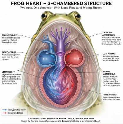
Let’s examine the heart. It is a muscular pumping structure located in the upper body cavity, protected by a membrane called the pericardium. Unlike our 4-chambered human heart, the frog heart has only three chambers: two atria at the top and a single ventricle at the bottom.
Deoxygenated blood from the major veins (vena cava) flows into a unique triangular structure called the sinus venosus, which attaches to the right atrium. After the blood is pumped through the ventricle, it exits into a sac-like structure on the ventral side called the conus arteriosus. From here, arteries distribute blood to the entire body (the arterial system), and veins return it back to the heart (the venous system).
Frogs also have special “short-cut” venous connections between specific organs. The connection between the liver and intestine is the hepatic portal system, while the connection between the kidneys and the lower body is the renal portal system.
What about the blood itself? Frog blood contains plasma, red blood cells (erythrocytes), white blood cells (leucocytes), and platelets. The red blood cells carry the red pigment haemoglobin for oxygen transport. Crucial difference: Unlike human mature RBCs, frog RBCs contain a nucleus! Lymph fluid is slightly different from blood; it lacks RBCs and certain proteins.
5.2 The Excretory System
To eliminate nitrogenous wastes, frogs use a highly developed excretory system comprising a pair of kidneys, ureters, the cloaca, and a urinary bladder.
The kidneys are dark red, compact, bean-like structures found a bit posteriorly in the body cavity, flanking the vertebral column. Inside these kidneys are the structural and functional filtering units known as uriniferous tubules or nephrons. Blood carries waste to the kidneys, where it is separated and excreted. Frogs primarily excrete urea, classifying them as ureotelic animals.
From the kidneys, tubes called ureters emerge. In male frogs, these ureters act as a shared pathway for both urine and sperm, making them a urinogenital duct that opens into the cloaca. In female frogs, however, the ureters and the oviduct open separately into the cloaca. The urinary bladder is thin-walled and sits just ventral to the rectum, eventually opening into the cloaca as well.
6. Nervous, Endocrine, and Reproductive Systems
6.1 Control and Coordination
To act quickly and catch flies, a frog needs stellar reflexes. This is managed by a highly evolved system involving both chemical (endocrine glands) and electrical (neural system) coordination.
- Endocrine Glands: These glands secrete chemical messengers called hormones. Major glands include the pituitary, thyroid, parathyroid, thymus, pineal body, pancreatic islets, adrenals, and gonads.
- Nervous System: This is divided into the Central Nervous System (CNS: brain and spinal cord), Peripheral Nervous System (PNS: cranial and spinal nerves), and Autonomic Nervous System (ANS: sympathetic and parasympathetic nerves). Notably, frogs have exactly ten pairs of cranial nerves originating from the brain.
The brain itself sits safely inside a bony brain box called the cranium. It is divided into three parts:
- Fore-brain: Contains olfactory lobes (for smell), paired cerebral hemispheres, and an unpaired diencephalon.
- Mid-brain: Features a distinct pair of optic lobes (for vision).
- Hind-brain: Composed of the cerebellum and medulla oblongata. The medulla exits the skull through a hole called the foramen magnum and continues down the vertebral column as the spinal cord.
6.2 Sense Organs
Frogs experience the world through several sensory avenues:
Sensory papillae for touch, taste buds for taste, nasal epithelium for smell, eyes for vision, and tympanum/internal ears for hearing. Out of these, only the eyes and internal ears are well-organized, distinct structures; the rest are basically clusters of cells clustered around nerve endings. Frog eyes are simple (one unit), spherical structures set in the skull’s orbits. They have no external ear flap—only the visible tympanum, which assists in both hearing and balancing (equilibrium).
6.3 The Reproductive System
Frogs possess distinct and well-organized male and female reproductive systems.
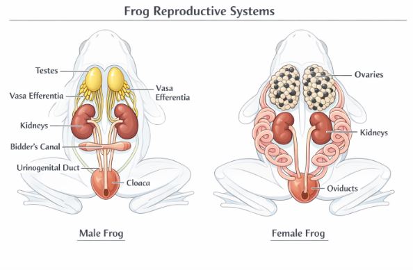
Male System: The male has a pair of yellowish, ovoid testes. These are glued to the upper section of the kidneys by a specialized double fold of tissue called the mesorchium. From the testes, about 10 to 12 tiny tubes called vasa efferentia arise, entering the kidneys and opening into a channel called Bidder’s canal. This canal finally merges with the urinogenital duct, which carries both urine and sperm out into the cloaca—a versatile median chamber used to pass feces, urine, and sperm outside the body.
Female System: The female features a pair of ovaries situated near the kidneys, but unlike males, there is no functional connection between her reproductive organs and her kidneys. A pair of oviducts carry eggs from the ovaries and open separately into the cloaca. A healthy, mature female can lay an astonishing 2500 to 3000 ova (eggs) at one time!
Because they are amphibians, fertilization is external—the male releases sperm over the eggs as the female lays them in the water. The eggs develop into a larval stage called a tadpole. Through a magnificent transformation process called metamorphosis, the aquatic tadpole eventually turns into a terrestrial adult frog.
7. Ecological and Economic Importance
Before we conclude, why should we care about frogs? Frogs are incredible friends to farmers and mankind because their diet consists heavily of insects, keeping pest populations down and protecting crops. Ecologically, they are a vital link in both the food chain and food web, helping maintain balance in our ecosystems. In some parts of the world, their highly muscular hind legs are even considered a culinary delicacy and used as food by humans.
Key Takeaways & Summary
- Cells form tissues, which form organs, which form organ systems to ensure survival through the division of labour.
- Epithelial tissues act as sheet-like linings for body surfaces, cavities, and ducts.
- Rana tigrina (Indian bullfrog) is cold-blooded, possesses a head and trunk (no neck/tail), and respires through its highly vascularized, mucous-covered skin in water .
- Digestive tract is short because they are carnivores. Liver and pancreas are the main digestive glands.
- Closed circulatory system with a 3-chambered heart and nucleated red blood cells.
- Excretory system utilizes kidneys and the cloaca. Males use a shared urinogenital duct.
- External fertilization occurs in water, leading to a tadpole stage that undergoes metamorphosis into an adult.
Common Student Misconceptions
Misconception 1: “Frogs drink a lot of pond water to stay hydrated.”
Correction: Absolutely false! Frogs never drink water through their mouths. They absorb all the necessary moisture directly through their specialized skin.
Misconception 2: “A frog’s ears look just like ours.”
Correction: Frogs completely lack an external ear structure (the pinna). The only visible part of their ear from the outside is the flat, membranous eardrum called the tympanum.
Practice Set: Test Your Knowledge (CBSE Pattern)
Very Short Answer Questions (1 Mark)
Q1. What is the scientific term used for the summer sleep exhibited by frogs?
Answer: The summer sleep undertaken by frogs to protect themselves from extreme heat is called aestivation.
Q2. Name the transparent structure that protects the frog’s eyes when it dives underwater.
Answer: The eyes are protected by a covering called the nictitating membrane.
Q3. State the primary nitrogenous waste excreted by frogs. Based on this, what type of animal is a frog?
Answer: Frogs excrete urea, making them ureotelic animals.
Short Answer Questions (2-3 Marks)
Q4. Explain the concept of sexual dimorphism in frogs. Provide specific examples.
Answer: Sexual dimorphism refers to the physical differences between male and female individuals of the same species. In frogs, males can be visually distinguished from females by the presence of sound-producing vocal sacs and a copulatory pad located on the first digit of their forelimbs. Both of these traits are absent in females.
Q5. Why is the alimentary canal of a frog relatively short compared to herbivores? Outline the path food takes from the stomach to the rectum.
Answer: The alimentary canal is short because frogs are carnivores, meaning their meat-based diet is easier and quicker to digest than plant matter. Food travels from the stomach as chyme into the duodenum, moves through the small intestine where final digestion and absorption occur, and the waste passes into the rectum before exiting through the cloaca .
Long Answer Questions (5 Marks)
Q6. Describe the structure and functioning of the blood vascular system in Rana tigrina. Highlight any unique structures in their heart and blood cells.
Answer: The frog possesses a well-developed, closed blood vascular system.
1. Heart Structure: The heart is a muscular organ located in the upper body cavity, wrapped in a pericardium. It has three chambers: two atria and one ventricle.
2. Key Vessels: A triangular sinus venosus joins the right atrium to receive blood from the major veins (vena cava). The ventricle pumps blood out into a sac-like conus arteriosus on the heart’s ventral side.
3. Portal Systems: Frogs feature special venous connections: the hepatic portal system (connecting liver and intestine) and the renal portal system (connecting kidneys and lower body).
4. Blood Composition: The blood consists of plasma, erythrocytes (RBCs), leucocytes (WBCs), and platelets. A unique feature of the frog’s red blood cells is that they are nucleated and contain the pigment haemoglobin.
Case-Based / Competency-Based Question (4 Marks)
Q7. Read the situation and answer the questions.
A group of students observed a frog sitting completely still in a deep mud burrow during the freezing month of January. It did not seem to be breathing through its mouth or nostrils, yet it was alive.
(a) What is the scientific term for this resting phase during winter?
(b) Why do frogs need to undergo this phase?
(c) Explain how the frog manages to survive without utilizing its lungs for respiration during this period.
Answer:
(a) This winter sleep phase is known as hibernation.
(b) Frogs are poikilotherms (cold-blooded), meaning they cannot regulate a constant internal body temperature. They hibernate in deep burrows to escape and survive extreme cold temperatures.
(c) During hibernation, the lungs are completely inactive. The frog relies entirely on cutaneous respiration, exchanging gases directly through its highly vascularized, moist skin.
Assertion-Reason Question
Q8. For the following question, two statements are given—one labeled Assertion (A) and the other labeled Reason (R). Select the correct answer from the codes (a), (b), (c), and (d) as given below.
(a) Both A and R are true, and R is the correct explanation of A.
(b) Both A and R are true, but R is not the correct explanation of A.
(c) A is true, but R is false.
(d) A is false, but R is true.
Assertion (A): The male frog utilizes the same tract to pass both urine and sperms into the cloaca.
Reason (R): In male frogs, the vasa efferentia open into Bidder’s canal, which eventually communicates with the urinogenital duct that comes out of the kidney.
Answer: (a). Both statements are true, and the anatomical connection (Reason) explaining how the reproductive tubes (vasa efferentia/Bidder’s canal) merge with the excretory tube perfectly explains why it is called a combined urinogenital duct in males .
End of Notes.
Students, make sure you practice drawing the neat diagrams of the digestive and reproductive systems of the frog, as these are high-yield topics for your exams! Happy studying!
Read Also:
Chapter 6- Anatomy of Flowering Plants
For official syllabus and textbooks, visit the
NCERT Official Website.