
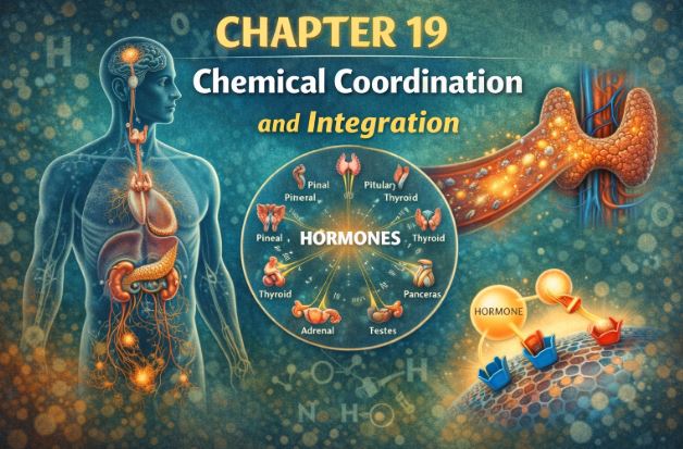
Introduction | Class 11 Biology Chapter 19
Hello students! Welcome back to another exciting chapter in Human Physiology. Today, we are going to talk about the body’s ultimate communication network. Have you ever wondered why your heart starts pounding instantly when a dog barks at you, but growing taller takes years? How does your body know exactly when to start digesting the food you just ate?
In our previous chapters, we discussed the nervous system. The neural system is like making a phone call or sending a direct text message: it is incredibly fast, specific, and point-to-point. However, it has a limitation. The nerve fibers cannot possibly reach every single tiny cell in your body, and a nerve impulse lasts only for a fraction of a second. But our body’s cells need continuous instructions to grow, metabolize, and survive.
This is where the Endocrine System steps in. Think of the endocrine system as a radio broadcast. It sends chemical messages out into the bloodstream, which then travel throughout the entire body. These messages are slow, sustained, and reach every corner of your system. Together, your nervous system and your endocrine system act as the joint CEOs of your body, coordinating and regulating every physiological function to keep you alive and healthy. Let’s dive into the fascinating world of hormones!
1. Endocrine Glands and Hormones: The Basics
1.1 What are Endocrine Glands?
In human biology, we generally have two types of glands. Exocrine glands have tiny tubes or “ducts” to pour their secretions (like sweat, saliva, or digestive enzymes) exactly where they are needed. Endocrine glands, on the other hand, are the rebels. They do not have ducts. Because they are “ductless glands,” they pour their magical chemicals directly into the blood surrounding them. The blood then acts as the delivery service, carrying these chemicals across the body.
1.2 Redefining Hormones
For a long time, we defined a hormone simply as a chemical produced by an endocrine gland, dumped into the blood, and sent to a distant target organ. But science is always evolving! Today, we have a much more accurate and scientific definition that you absolutely must memorize for your exams.
Modern Definition of a Hormone: Hormones are non-nutrient chemicals that act as intercellular messengers (messengers between cells) and are produced in trace (very tiny) amounts.
Why this new definition? Because we now know that it isn’t just the main endocrine glands that make hormones. Even your heart, kidneys, and stomach line secrete chemical messengers! While lower animals like insects have very simple endocrine setups, vertebrates (like us humans) have a beautifully complex system with a wide array of chemicals ensuring smooth internal coordination.
2. The Human Endocrine System: A Tour of Your Body
Imagine a map of your body. Scattered from your brain down to your lower abdomen are special groups of cells and tissues dedicated solely to hormone production. The primary organized endocrine bodies include the Hypothalamus, Pituitary, Pineal, Thyroid, Parathyroid, Thymus, Adrenal glands, Pancreas, and the Gonads (Testes in males and Ovaries in females). Let’s study them one by one, starting from the top of your head!
)
2.1 The Hypothalamus: The Supreme Commander
Located at the base of the diencephalon in your forebrain, the hypothalamus is the true master of your internal environment. It bridges the gap between your nervous system and your endocrine system. Inside the hypothalamus are clusters of specialized neurons called nuclei. These aren’t regular nerve cells; they are neurosecretory cells, meaning they secrete hormones!
The hypothalamus has one main job: controlling the pituitary gland. It produces two categories of hormones:
- Releasing Hormones: These act as a green light, telling the pituitary to release its own hormones. Example: Gonadotrophin Releasing Hormone (GnRH) tells the pituitary to release hormones that act on the gonads.
- Inhibiting Hormones: These act as a red light, telling the pituitary to stop. Example: Somatostatin is sent down to tell the pituitary to stop releasing Growth Hormone.
Teacher’s Note: The hypothalamus connects to the front part of the pituitary via a special blood vessel system called the portal circulatory system. But it connects to the back part of the pituitary directly through nerve fibers!
2.2 The Pituitary Gland: The Master Gland
The pituitary is a tiny, pea-sized gland sitting safely inside a bony cavity in your skull known as the sella tursica. It hangs right below the hypothalamus by a stalk. Anatomically, it is split into two main sections: the Anterior Pituitary (Adenohypophysis) and the Posterior Pituitary (Neurohypophysis).
A. Anterior Pituitary (Pars Distalis)
This section is a hormone factory. It manufactures and releases several major hormones:
- Growth Hormone (GH): Regulates your body’s growth. If a child makes too much GH, they grow exceptionally tall (Gigantism). If they make too little, it leads to stunted growth (Pituitary Dwarfism). In adults, excess GH causes a severe disfiguring disease called Acromegaly, which enlarges the face and hands and can be life-threatening if ignored.
- Prolactin (PRL): Responsible for the growth of mammary glands and the production of milk in females.
- Thyroid Stimulating Hormone (TSH): Travels to your neck to tell the thyroid gland to get to work.
- Adrenocorticotrophic Hormone (ACTH): Travels to the adrenal glands to stimulate the release of steroid hormones called glucocorticoids.
- Gonadotrophins (LH and FSH): Luteinizing Hormone and Follicle Stimulating Hormone target the reproductive organs. In males, they drive sperm production and testosterone release. In females, they control the menstrual cycle, ovulation of the Graafian follicle, and maintaining the corpus luteum.
Note on Pars Intermedia: In humans, this middle part is almost merged with the anterior pituitary. It produces just one hormone, Melanocyte Stimulating Hormone (MSH), which acts on skin cells to regulate pigmentation.
B. Posterior Pituitary (Pars Nervosa)
Here is a massive trick question examiners love: Which hormones are synthesized by the posterior pituitary? The answer is NONE! The posterior pituitary only stores and releases hormones that were actually manufactured up in the hypothalamus and transported down through nerve axons.
- Oxytocin: The “cuddle hormone” or “birth hormone.” It causes strong smooth muscle contractions. In females, it is responsible for the massive uterine contractions during childbirth and the ejection of milk from mammary glands.
- Vasopressin (Anti-diuretic Hormone – ADH): This acts on your kidneys. It tells the distal tubules of the kidney to reabsorb water back into the blood, preventing you from peeing out all your body’s water (diuresis). If your body fails to make ADH, you suffer from constant urination and severe dehydration, a dangerous condition called Diabetes Insipidus.
)
3. The Pineal and Thyroid Glands
3.1 The Pineal Gland: Your Biological Clock
Tucked away on the dorsal (back) side of your forebrain is the tiny pineal gland. Its main product is a hormone called Melatonin. Have you ever experienced jet lag after a long flight? That’s your melatonin getting confused! Melatonin is the chief regulator of your 24-hour diurnal rhythm (circadian rhythm). It manages your sleep-wake cycle and daily body temperature changes. It also has subtle influences on your metabolism, skin pigmentation, menstrual cycles, and even your immune defense.
3.2 The Thyroid Gland: The Metabolism Engine
Wrap your fingers around the front of your neck, just below the Adam’s apple. That’s where your butterfly-shaped thyroid gland lives. It has two lobes resting on either side of your windpipe (trachea), connected by a thin band of tissue called the isthmus.
Inside, the gland is made of tiny follicles. These follicles pull iodine from your blood to manufacture two critical hormones: Tetraiodothyronine (Thyroxine or T4) and Triiodothyronine (T3).
Functions of Thyroid Hormones:
- They control your Basal Metabolic Rate (BMR) – the speed at which your body burns fuel at rest.
- They support the creation of red blood cells.
- They manage how you metabolize carbohydrates, proteins, and fats.
- They help maintain water and electrolyte balance.
The thyroid also makes a non-iodine protein hormone called Thyrocalcitonin (TCT), which lowers calcium levels in the blood.
Thyroid Disorders
If your diet lacks iodine, your thyroid struggles to make hormones. To compensate, the gland swells up massively, a condition known as Goitre (Hypothyroidism). If a pregnant woman has hypothyroidism, it is disastrous for the fetus, causing stunted growth, severe mental retardation, and a condition called Cretinism.
Conversely, sometimes the gland goes into overdrive, often due to cancer or nodules, releasing too much hormone. This is Hyperthyroidism. A famous form of this is Graves’ disease (Exopthalmic goitre), where a person suffers from rapid weight loss, an abnormally high metabolic rate, a swollen neck, and distinctively bulging eyeballs.
4. Parathyroid and Thymus Glands
4.1 Parathyroid Glands: The Bone Miners
Hidden on the back surface of the thyroid gland are four tiny dots called the parathyroid glands. They secrete a single, incredibly important peptide called Parathyroid Hormone (PTH).
PTH is the ultimate protector of your blood calcium levels. Calcium isn’t just for strong bones; it is essential for your heart to beat and your nerves to fire. If your blood calcium drops, PTH is released. It acts like a miner, going to your bones and breaking them down (demineralization) to release calcium into the blood. It also tells the kidneys to save calcium from urine and the intestines to absorb more calcium from your food. Because it raises blood calcium, we call PTH a hypercalcemic hormone. It works perfectly opposite to the thyroid’s TCT.
4.2 Thymus: The Immune Training Academy
Located right behind your breastbone (sternum) and between your lungs is a lobular gland called the thymus. This gland is the boot camp for your immune system. It secretes peptide hormones called Thymosins.
Thymosins are critical for the maturation of T-lymphocytes, the special white blood cells that provide cell-mediated immunity (attacking infected cells directly). They also help promote antibodies for humoral immunity.
Fascinating Fact: The thymus is large and active in children but slowly shrinks and degenerates as we grow old. This is exactly why elderly people have weaker immune responses and fall sick more easily!
5. Adrenal Glands: The Stress Managers
Sitting like little triangular hats on top of both your kidneys are the adrenal glands. Think of the adrenal gland like a chocolate truffle: it has an outer shell (the cortex) and an inner filling (the medulla). They do completely different jobs.
)
5.1 The Adrenal Medulla (The Inner Core)
This area is responsible for your “Fight or Flight” response. When you are terrified, stressed, or in an emergency, the medulla instantly pumps out Adrenaline (Epinephrine) and Noradrenaline (Norepinephrine). These are known as catecholamines.
Within seconds of seeing a threat, these hormones cause your pupils to dilate, your hair to stand on end (piloerection), and you to start sweating. They make your heart beat faster and stronger, and increase your breathing rate. To give your muscles energy to run or fight, they break down stored glycogen into glucose, flooding your blood with sugar.
5.2 The Adrenal Cortex (The Outer Shell)
The cortex is divided into three zones (outer glomerulosa, middle fasciculata, inner reticularis) and produces essential steroid hormones called corticoids.
- Glucocorticoids (Mainly Cortisol): These manage carbohydrate metabolism. Cortisol helps you deal with long-term stress. It stimulates gluconeogenesis (making new glucose), breaks down fats and proteins, and actively suppresses the immune system. This is why doctors use cortisol creams to stop extreme inflammatory allergic reactions!
- Mineralocorticoids (Mainly Aldosterone): These control water and salt balance. Aldosterone tells your kidneys to hold onto Sodium ($Na^+$) and water, while flushing out Potassium ($K^+$). This keeps your blood pressure and fluid volume stable.
- Androgenic Steroids: Small amounts of sex hormones are made here, contributing to the growth of pubic, facial, and axial hair during puberty.
If the adrenal cortex gets damaged and underproduces hormones, carbohydrate metabolism crashes, leading to severe weakness and fatigue. This dangerous condition is called Addison’s disease.
6. The Pancreas: The Sugar Seesaw
The pancreas is a unique “composite” gland. This means it acts as an exocrine gland (pouring digestive juices into the intestine) AND an endocrine gland. The endocrine portion consists of tiny clusters of cells scattered like islands, beautifully named the Islets of Langerhans. There are about 1 to 2 million of these islands, but they make up barely 1-2% of the whole pancreas.
Inside these islets are two VIP cells playing a constant game of tug-of-war with your blood sugar:
- Alpha ($\alpha$) cells secrete Glucagon: Imagine you haven’t eaten for 10 hours. Your blood sugar is dangerously low. Glucagon is released, acting on your liver cells (hepatocytes). It forces the liver to break down stored glycogen into glucose and dump it into the blood. Therefore, glucagon is a hyperglycemic hormone.
- Beta ($\beta$) cells secrete Insulin: You just ate a massive bowl of rice. Your blood sugar spikes. Insulin is released. It acts like a key, unlocking your body’s cells (especially liver and fat cells) so they can absorb glucose from the blood and use it or store it. Because it lowers blood sugar, insulin is a hypoglycemic hormone.
The Crisis of Diabetes Mellitus:
If your pancreas fails to produce enough insulin, or your cells become resistant to it, glucose cannot enter your cells. It builds up to toxic levels in the blood, leading to a complex disorder called Diabetes Mellitus. The body starts desperately flushing the excess sugar out through urine. Starved of glucose, cells start burning fat incorrectly, creating harmful chemical byproducts called ketone bodies. Today, we successfully treat this condition using synthetic insulin therapy.
7. Gonads (Testes and Ovaries)
While their main job is to produce gametes (sperm and eggs), the gonads are also powerful endocrine glands.
7.1 Testis (Male)
Located outside the abdomen in the scrotal sac, the testes contain seminiferous tubules and interstitial spaces. Specialized cells called Leydig cells sit in these spaces and pump out androgens, primarily Testosterone.
Testosterone is responsible for male puberty. It develops the male accessory organs, triggers muscle growth, deepens the voice, grows facial hair, drives aggressive behavior, and regulates male sexual desire (libido). Most importantly, it is the crucial stimulator for making sperm (spermatogenesis).
7.2 Ovary (Female)
Located inside the abdomen, the ovaries produce a single egg every month. They secrete two major steroid hormones:
- Estrogen: Produced by growing ovarian follicles. It manages the development of female secondary sex characters (like breast development and a high-pitched voice), regulates female sexual behavior, and thickens the uterine lining.
- Progesterone: Once an egg is released, the broken follicle shell turns into a yellow body called the corpus luteum. This structure pumps out progesterone, the ultimate “pregnancy hormone.” It supports the pregnancy and stimulates the mammary glands to form milk-storing sacs (alveoli).
8. Hormones from Non-Endocrine Tissues
Remember how we said the definition of hormones changed? That’s because other organs pitch in too!
- The Heart: If your blood pressure gets too high, the walls of the heart’s atria stretch and release a peptide called Atrial Natriuretic Factor (ANF). ANF causes blood vessels to dilate (widen), which beautifully reduces blood pressure.
- The Kidneys: Special Juxtaglomerular cells in the kidney sense low oxygen and release Erythropoietin. This hormone travels to bone marrow and stimulates the creation of brand new Red Blood Cells (erythropoiesis).
- The Gastrointestinal (GI) Tract: Your stomach and intestines secrete four main local hormones to manage digestion:
- Gastrin: Tells the stomach to release acid and pepsinogen.
- Secretin: Tells the pancreas to release water and bicarbonates.
- Cholecystokinin (CCK): Squeezes the gallbladder for bile and the pancreas for enzymes.
- Gastric Inhibitory Peptide (GIP): Acts as the brakes, stopping gastric secretion when food moves on.
9. Mechanism of Hormone Action (How do they actually work?)
This is a highly conceptual topic. Let’s break it down using a simple daily life analogy!
A hormone floating in the blood passes by billions of cells, but it only affects its specific target cell. Why? Because target cells have specific 3D protein structures called Hormone Receptors. It works exactly like a lock and a key. If the hormone (key) doesn’t fit the receptor (lock), nothing happens. When they do connect, they form a hormone-receptor complex.
Hormones fall into different chemical classes (Proteins/peptides, Steroids, Iodothyronines, and Amino-acid derivatives). Depending on their chemistry, they interact with cells in two entirely different ways:
Type A: The Doorbell Method (Water-Soluble / Protein Hormones)
Protein hormones (like Insulin, Glucagon, Pituitary hormones) are water-soluble. They cannot push through the oily lipid membrane of a cell.
How it works: The hormone arrives at the cell membrane and binds to a Membrane-Bound Receptor on the outside surface. It acts like a person ringing a doorbell. The hormone never actually enters the cell! Pressing the “doorbell” triggers a series of events inside the cell that generate a Second Messenger (like Cyclic AMP, $IP_{3}$, or $Ca^{2+}$). This second messenger runs around inside the cell, altering enzymes and changing the cell’s metabolism to create the desired physiological response.
)
Type B: The VIP Guest Method (Lipid-Soluble / Steroid Hormones)
Steroid hormones (like Cortisol, Testosterone, Estrogen) and Thyroid hormones are lipid-soluble. They easily melt right through the cell’s fatty membrane.
How it works: They do not need a doorbell. They walk straight inside the cell and bind to an Intracellular Receptor, usually located deep inside the nucleus. The hormone-receptor complex attaches directly to the cell’s DNA (genome). This interaction turns specific genes on or off, creating new messenger RNA (mRNA) and ultimately new proteins. Over time, these cumulative biochemical changes result in tissue growth and major physiological changes.
)
Key Takeaways & Summary
- The endocrine system provides slow but widespread and sustained chemical coordination using hormones.
- Hormones are trace-amount, non-nutrient intercellular messengers.
- The Hypothalamus controls the anterior pituitary via releasing/inhibiting hormones, and stores oxytocin/ADH in the posterior pituitary.
- The Thyroid regulates metabolism (needs iodine), while Parathyroid controls blood calcium levels.
- The Adrenal gland has two parts: Medulla handles emergency stress (fight or flight), and Cortex handles long-term stress and salt balance.
- The Pancreas maintains blood sugar via Glucagon (raises) and Insulin (lowers).
- Hormones work either by binding to membrane receptors (creating second messengers) or by entering the cell to interact with DNA.
Common Student Misconceptions
Misconception 1: The posterior pituitary manufactures Oxytocin and ADH.
Correction: Absolute trap! The posterior pituitary is just a storage warehouse. The actual manufacturing plant for these hormones is the hypothalamus.
Misconception 2: Diabetes Mellitus and Diabetes Insipidus are the same disease.
Correction: They share the word “Diabetes” (meaning heavy urination) but are totally different. Diabetes Mellitus is a sugar problem (lack of insulin from the pancreas). Diabetes Insipidus is a water problem (lack of ADH from the hypothalamus/pituitary), where the urine has no sugar but you lose dangerous amounts of water.
Practice Set: Test Your Knowledge (CBSE Pattern)
Very Short Answer Questions (1 Mark)
Q1. Why is the parathyroid hormone considered a hypercalcemic hormone?
Answer: PTH is called a hypercalcemic hormone because its primary action is to increase the concentration of calcium ions ($Ca^{2+}$) in the blood by triggering bone demineralization and kidney reabsorption.
Q2. Name the hormone that regulates the 24-hour diurnal rhythm of our body. Where is it secreted from?
Answer: Melatonin. It is secreted by the pineal gland located on the dorsal side of the forebrain.
Q3. Which endocrine gland is large at the time of birth but reduces in size with age?
Answer: The Thymus gland.
Short Answer Questions (3 Marks)
Q4. Differentiate between the mechanisms of action of a protein hormone and a steroid hormone.
Answer:
1. Receptor Location: Protein hormones bind to membrane-bound receptors on the target cell surface. Steroid hormones pass through the membrane and bind to intracellular receptors inside the nucleus.
2. Second Messengers: Protein hormones generate second messengers (like cyclic AMP or $Ca^{2+}$) to alter cell metabolism. Steroid hormones do not require second messengers.
3. Cellular Action: Protein hormones generally activate existing enzymes for rapid physiological responses. Steroid hormones interact with the cell’s genome to regulate gene expression and synthesize new proteins, taking a longer time.
Q5. Write short notes on the functions of (a) Insulin (b) Aldosterone.
Answer:
(a) Insulin: Secreted by beta-cells of the pancreas, it regulates glucose homeostasis. It enhances cellular uptake of glucose from the blood by hepatocytes and adipocytes, lowering blood sugar (hypoglycemia). It also promotes the conversion of glucose to glycogen.
(b) Aldosterone: A mineralocorticoid secreted by the adrenal cortex. It acts on kidney tubules to stimulate the reabsorption of sodium ions and water, and the excretion of potassium ions. This maintains body fluid volume, electrolytes, and blood pressure.
Long Answer Questions (5 Marks)
Q6. Discuss the hormones secreted by the anterior pituitary and describe their specific functions. Name the disorders caused by the over and under-secretion of Growth Hormone.
Answer: The anterior pituitary (pars distalis) secretes the following crucial trophic hormones:
1. Growth Hormone (GH): Regulates general body growth and metabolism.
2. Prolactin (PRL): Stimulates the growth of mammary glands and milk formation.
3. Thyroid Stimulating Hormone (TSH): Stimulates the thyroid gland to synthesize and release thyroid hormones.
4. Adrenocorticotrophic Hormone (ACTH): Stimulates the adrenal cortex to release glucocorticoids like cortisol.
5. Luteinizing Hormone (LH): In males, it stimulates testes to produce androgens. In females, it induces ovulation of mature follicles and maintains the corpus luteum.
6. Follicle Stimulating Hormone (FSH): Regulates spermatogenesis in males and stimulates the growth of ovarian follicles in females.
Disorders of GH:
– Over-secretion in childhood causes Gigantism.
– Under-secretion in childhood causes Pituitary Dwarfism.
– Over-secretion in adults causes Acromegaly (severe disfigurement of the face and extremities).
Case-Based / Competency-Based Question (4 Marks)
Q7. Read the situation and answer the questions.
A 45-year-old patient named Raj complains of extreme fatigue, constant hunger, excessive thirst, and frequent urination. Blood tests reveal an abnormally high level of glucose in his blood, and a urine test shows the presence of glucose and ketone bodies. Based on this clinical picture:
(a) Identify the complex disorder Raj is suffering from.
(b) Which hormone is deficient or ineffective in his body, and which specific cells secrete it?
(c) Contrast this condition with another disorder where a patient experiences excessive urination but no glucose is lost in the urine.
Answer:
(a) Raj is suffering from Diabetes Mellitus, characterized by prolonged hyperglycemia and loss of glucose via urine.
(b) The hormone Insulin is deficient or ineffective. It is secreted by the beta ($\beta$) cells of the Islets of Langerhans in the pancreas.
(c) The contrasting condition is Diabetes Insipidus. It involves excessive water loss and urination due to a deficiency of Anti-Diuretic Hormone (ADH/Vasopressin) from the posterior pituitary, but the patient’s blood sugar levels remain completely normal and no glucose is found in the urine.
Assertion-Reason Question
Q8. For the following question, two statements are given—one labeled Assertion (A) and the other labeled Reason (R). Select the correct answer from the codes (a), (b), (c), and (d) as given below.
(a) Both A and R are true, and R is the correct explanation of A.
(b) Both A and R are true, but R is not the correct explanation of A.
(c) A is true, but R is false.
(d) A is false, but R is true.
Assertion (A): Adrenaline is famously called the emergency hormone or the hormone of “Fight or Flight.”
Reason (R): Adrenaline acts on the liver to stimulate glycogenesis (conversion of glucose to glycogen), thus storing energy during stress.
Answer: (c) A is true, but R is false.
Explanation: The assertion is perfectly correct. Adrenaline is released during stress. However, the reason is entirely false. Adrenaline stimulates glycogenolysis (breaking down glycogen INTO glucose) to increase blood sugar levels, providing instant, available energy for the muscles to fight or run. It does not store energy during emergencies; it burns it!
End of Notes.
Students, remember that physiology is all about connections. Don’t just memorize the hormones; understand the “why” and “where” of each chemical signal. Draw out the hormone pathways on a blank sheet of paper for the best revision. Happy studying!
Read Also:
Class-11 Biology All Chapters
For official syllabus and textbooks, visit the
NCERT Official Website.