
Introduction | Class 11 Biology Excretory Products Notes
Welcome back, future doctors and biologists! Today, we are exploring one of the most brilliant and under-appreciated systems in the human body. Think about your house for a moment. You bring in groceries, cook meals, and consume things daily. But what happens to the wrappers, the vegetable peels, and the leftovers? They go into the trash, and eventually, the garbage truck takes them away. If the garbage stays inside, the house becomes toxic. Your body works exactly the same way!
Every single second, your cells are performing thousands of metabolic reactions. While these reactions produce the energy and materials you need to live, they also generate waste. Substances like carbon dioxide, water, various ions, and specifically, nitrogen-containing wastes must be removed[cite: 11, 12]. If they accumulate, they can severely damage your cells. In this chapter, we will uncover how our bodies act as master filters, sorting the good from the bad, and safely eliminating the dangerous toxins while holding onto the life-saving water and nutrients. Let us dive into the fascinating world of excretion!
1. The Big Three: Types of Nitrogenous Wastes
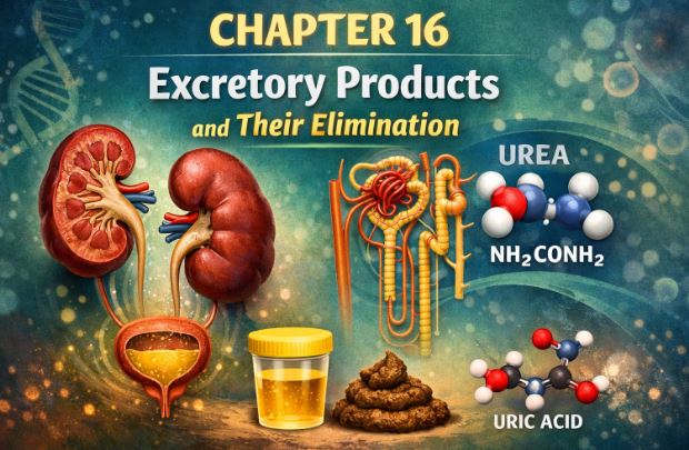
When we break down proteins and nucleic acids in our food, our bodies produce nitrogen-based waste. Depending on the animal and where it lives, this waste is thrown out in one of three major forms: Ammonia, Urea, or Uric Acid[cite: 13]. The environment an animal lives in usually dictates which waste product it produces[cite: 326].
1.1 Ammonotelism (Excreting Ammonia)
Ammonia is extremely toxic[cite: 14]. Think of it as a highly corrosive chemical. Because it is so dangerous, it cannot be stored in the body for long and needs a massive amount of water to be diluted and flushed out[cite: 14].
Therefore, animals that live in water, like bony fishes, aquatic insects, and aquatic amphibians, naturally excrete ammonia[cite: 15]. Because ammonia is highly soluble, fishes don’t even need complex kidneys for this; they simply let it diffuse out through their gills or body surface straight into the surrounding water[cite: 16, 17].
1.2 Ureotelism (Excreting Urea)
Now, what about land animals like us? We cannot afford to lose gallons of water just to flush out ammonia. So, land adaptation forced us to create a less toxic waste product: Urea[cite: 18]. Mammals (including humans), marine fishes, and many terrestrial amphibians are ureotelic[cite: 19].
Here is how it works: Our liver takes the highly toxic ammonia and chemically converts it into the much safer urea[cite: 20]. The blood carries this urea to the kidneys, where it is filtered out and removed from the body[cite: 20].
1.3 Uricotelism (Excreting Uric Acid)
Some animals live in extremely dry conditions or need to be very light for flight. They cannot afford to lose almost any water. Birds, reptiles, land snails, and insects convert their waste into uric acid[cite: 22]. Uric acid is the least toxic of the three and can be excreted as a thick white paste or pellet, conserving a tremendous amount of water[cite: 14, 22]. Have you ever noticed bird droppings? That white, chalky substance is uric acid!
2. A Quick Tour of the Animal Kingdom’s Filters
Before we zoom in on humans, let’s appreciate how other creatures handle their waste. Nature has designed various filtering structures:
- Protonephridia (Flame Cells): Found in simple creatures like flatworms (Planaria), rotifers, and Amphioxus. These structures mainly handle osmoregulation, which means keeping the water and salt balance perfectly tuned[cite: 34, 35].
- Nephridia: If you dig up an earthworm, it uses these tiny tubular structures to balance its body fluids and remove nitrogenous waste[cite: 36, 37].
- Malpighian Tubules: Found in most insects like the cockroach scurrying across the floor. They handle both waste removal and osmoregulation[cite: 38, 39].
- Antennal Glands (Green Glands): Found in crustaceans like prawns[cite: 40].
3. The Human Excretory System
This is the core of our chapter. Get ready to understand the incredible plumbing of your own body.
Our excretory system consists of a team of organs: one pair of kidneys, one pair of ureters, a single urinary bladder, and a urethra[cite: 54].

3.1 The Kidneys: Your Built-in Purifiers
Your kidneys are two reddish-brown, bean-shaped organs[cite: 55]. They are located near your back, roughly between the last thoracic and third lumbar vertebrae[cite: 55]. Despite doing a massive job, an adult kidney is quite small—about 10-12 cm long and weighing around 120-170 grams[cite: 56].
If you cut a kidney open, you’ll see a specific internal architecture. The outer part is called the cortex, and the inner part is the medulla[cite: 60]. The medulla is divided into cone-like structures called medullary pyramids[cite: 61]. The cortex doesn’t just stay on the outside; it pushes down between these pyramids to form columns known as the Columns of Bertini[cite: 62, 66].
On the inner curve of the kidney, there is a notch called the hilum[cite: 57]. Think of the hilum as the gateway where the renal artery (bringing dirty blood in), the renal vein (taking clean blood out), and the ureter (taking urine away) all connect[cite: 57]. Inner to this gateway is a broad, funnel-like space called the renal pelvis[cite: 58, 59].
3.2 The Nephron: The Microscopic Marvel
The kidney is not a hollow bag; it is densely packed with nearly one million microscopic filters called Nephrons[cite: 68]. The nephron is the structural and functional unit of the kidney[cite: 68]. Each nephron has two main components: the Glomerulus and the Renal Tubule[cite: 69].

1. The Glomerulus: This is a highly tangled ball of blood capillaries. Blood enters this ball through the afferent arteriole and leaves through a narrower vessel called the efferent arteriole[cite: 70, 71].
2. The Renal Tubule: This long tube catches the filtered fluid and processes it. It starts with a cup-shaped structure called Bowman’s capsule that perfectly wraps around the glomerulus[cite: 72]. Together, the glomerulus and Bowman’s capsule are referred to as the Malpighian body[cite: 73].
From the Bowman’s capsule, the tube twists and turns. Here is the sequence of the journey:
- Proximal Convoluted Tubule (PCT): The highly coiled first section[cite: 74].
- Loop of Henle: A hairpin-like loop dipping down into the kidney. It has a descending limb going down, and an ascending limb coming back up[cite: 105].
- Distal Convoluted Tubule (DCT): Another coiled section after the loop[cite: 106].
- Collecting Duct: The final straight tube where many nephrons pour their processed fluid, eventually leading to the renal pelvis[cite: 107].
4. The Process of Urine Formation
Forming urine isn’t just about dumping water. It is a highly calculated 3-step process: Glomerular filtration, Reabsorption, and Secretion[cite: 117].
Step 1: Glomerular Filtration (Ultrafiltration)
Blood rushes into the glomerulus under high pressure. This pressure forces the liquid part of the blood through three very thin layers into the Bowman’s capsule[cite: 119, 121]. The inner lining cells of the Bowman’s capsule, called podocytes, are arranged intricately to leave tiny “filtration slits”[cite: 122].
This filter is so incredibly fine that almost everything in the blood plasma (water, glucose, salts, urea) passes through, except large proteins and blood cells[cite: 123]. Because of this fine precision, we call it ultrafiltration[cite: 124].
Teacher’s Fact: Your kidneys filter an astonishing 180 liters of fluid a day! We call this the Glomerular Filtration Rate (GFR)[cite: 128, 129].
Step 2: Selective Reabsorption
Wait, if we filter 180 liters a day, why do we only urinate about 1.5 liters? Because 99% of what was filtered is extremely valuable and must be taken back into the blood[cite: 134]. This taking-back process is called reabsorption[cite: 135].
The tubules pull back the good stuff (like 100% of the glucose and amino acids) using active transport, while water simply follows along passively[cite: 136, 137].
Step 3: Tubular Secretion
Sometimes, harmful items or excess ions escape the first filtration step. The cells lining the tubule actively pick up these hidden wastes (like excess potassium, hydrogen ions, and ammonia) from the surrounding blood vessels and secrete them into the tubule fluid[cite: 139]. This final step is crucial for maintaining the acid-base (pH) balance of our bodily fluids[cite: 140].
5. Functions of the Tubules
Let’s look at exactly what happens in each part of the nephron’s tubing.
- PCT: This is the heavy lifter. Its walls have brush borders to increase surface area[cite: 144]. It reabsorbs nearly all essential nutrients and 70-80% of water and electrolytes[cite: 145].
- Henle’s Loop: This part acts like a salt and water separator. The descending limb lets water exit but blocks salts[cite: 148]. So, the fluid gets highly concentrated. Then, it turns the corner. The ascending limb blocks water but actively pumps out salts[cite: 149]. This makes the fluid diluted again[cite: 150].
- DCT: This is the fine-tuning area. Depending on what the body needs, it performs conditional reabsorption of water and sodium[cite: 151]. It also secretes hydrogen and potassium to keep blood pH stable[cite: 152].
- Collecting Duct: This long tube runs deep into the kidney’s medulla[cite: 178]. Here, massive amounts of water can be squeezed out of the fluid and reabsorbed, creating highly concentrated urine[cite: 179].
6. The Counter-Current Mechanism: Concentrating the Urine
This is a favorite topic for examiners, so read carefully! How does a desert-dwelling mammal or a human survive without drinking water constantly? We make concentrated urine.

Wrapped around the Loop of Henle is a special U-shaped blood vessel called the Vasa recta[cite: 114]. The fluid inside the Loop of Henle flows in one direction, while the blood in the Vasa recta flows in the exact opposite direction. This opposing flow creates a “counter-current”[cite: 186, 187].
By working together, these two U-shaped tubes trap salt (NaCl) and urea in the deep inner tissue (interstitium) of the kidney medulla[cite: 191]. They create a steep concentration gradient—it goes from a normal 300 mOsmolL⁻¹ near the top to a super-salty 1200 mOsmolL⁻¹ at the very bottom[cite: 190].
Why do we want the inner kidney to be so salty? Because when the final collecting duct carries urine through this salty deep zone, the salt draws water out of the collecting duct[cite: 258]. The water is saved and put back into the blood, leaving behind a highly concentrated urine—up to four times more concentrated than the initial filter fluid! [cite: 259]
7. Regulation of Kidney Function: The Management Team
Your kidneys don’t work blindly. They are managed by hormonal signals based on how much water is in your body.
7.1 The Hypothalamus and ADH
If you sweat a lot and lose body fluid, sensors called osmoreceptors detect this drop[cite: 262]. They tell a brain region called the hypothalamus to release a hormone named Antidiuretic Hormone (ADH), also known as vasopressin[cite: 263]. ADH rushes to the kidneys and forces the later parts of the tubules to reabsorb lots of water, preventing you from losing more fluid via urine (diuresis)[cite: 264]. If you drink plenty of water, ADH release stops, and you produce more urine[cite: 265].
7.2 The RAAS System (Renin-Angiotensin)
If your blood pressure drops sharply, the kidneys sense it using a special area called the Juxtaglomerular Apparatus (JGA)[cite: 268]. The JGA cells release an enzyme called Renin[cite: 268]. Renin acts like a domino effect, eventually producing a powerful chemical called Angiotensin II[cite: 268]. Angiotensin II squeezes your blood vessels to raise blood pressure and tells the adrenal glands to release Aldosterone[cite: 269, 270]. Aldosterone forces the kidneys to reabsorb salt and water, further boosting blood pressure[cite: 271, 272].
7.3 The Heart’s Brake Pedal: ANF
If blood pressure gets too high, the heart gets stretched. The heart cells release Atrial Natriuretic Factor (ANF)[cite: 274]. ANF dilates the blood vessels, acting as a direct check to slow down the RAAS system and reduce blood pressure[cite: 275, 276].
8. Micturition (The Release of Urine)
Once urine is fully formed, it trickles down the ureters and fills the urinary bladder[cite: 279]. As the bladder fills up, its walls stretch. This stretching activates receptors that send an urgent signal to your Central Nervous System (CNS)[cite: 280, 281].
When the time is right, your brain sends a motor signal back. It commands the bladder muscles to contract and simultaneously tells the urethral sphincter (a muscular valve) to relax, allowing urine to flow out[cite: 287]. This reflex action is known as Micturition[cite: 288].
9. Other Organs That Help in Excretion
The kidneys aren’t alone in the waste disposal business!
- Lungs: Every time you exhale, you remove huge amounts of toxic carbon dioxide (about 200mL per minute) and water vapor[cite: 296].
- Liver: The largest gland in your body. It dumps old cholesterol, degraded hormones, and byproducts of broken-down red blood cells (like bilirubin) into the bile, which leaves the body via your digestive waste[cite: 297, 298].
- Skin: Sweat glands remove water, salt, and small amounts of urea, mainly to cool you down[cite: 300]. Sebaceous (oil) glands remove certain sterols and waxes, providing a protective covering for your skin[cite: 301, 302].
10. Disorders of the Excretory System
When the system breaks down, serious problems arise:
- Uremia: If kidneys malfunction, urea builds up in the blood to dangerous levels. This can be fatal[cite: 306].
- Hemodialysis: An artificial kidney machine used for uremic patients. Blood is pumped into a machine containing a cellophane tube surrounded by a special fluid[cite: 308, 310]. Wastes diffuse out of the blood into the fluid, and clean blood is returned to the body[cite: 315, 316].
- Renal Calculi: Commonly known as kidney stones. These are solid, crystallized masses of salts (like oxalates) formed inside the kidney, causing extreme pain[cite: 321].
- Glomerulonephritis: An inflammatory condition affecting the delicate glomeruli filters within the kidneys[cite: 322].
Real-Life Examples to Understand Excretion
- The Coffee Filter Analogy: Think of the glomerulus and Bowman’s capsule like making drip coffee. The coffee filter (glomerulus) lets the water and flavor molecules (plasma, glucose, small salts) pass through into the pot, but blocks the large, chunky coffee grounds (red blood cells and large proteins) from getting in.
- The Factory Recycling Plant: Reabsorption is exactly like a recycling center at a factory. Even though the factory throws out 180 boxes a day into the sorting bin, the smart workers (tubules) realize that 99% of those boxes contain useful parts (glucose, water). They grab those useful boxes off the conveyor belt and put them back inside the factory, leaving only the real trash (urea) to be thrown away.
Key Takeaways & Summary
- Animals excrete nitrogenous wastes based on water availability: Ammonia (aquatic), Urea (mammals/amphibians), Uric Acid (birds/reptiles)[cite: 326, 327].
- The nephron is the functional unit of the kidney, consisting of the glomerulus and the renal tubule[cite: 332].
- Urine formation includes ultrafiltration, selective reabsorption (mostly in PCT), and tubular secretion[cite: 337].
- The Loop of Henle and Vasa recta use a counter-current mechanism to create a salty medullary gradient, allowing us to produce highly concentrated urine and save water[cite: 348, 349].
- Kidney function is heavily regulated by hormones like ADH (saves water) and the RAAS system (regulates blood pressure).
Common Student Misconceptions
Misconception 1: Sweat is primarily an excretory mechanism.
Correction: While sweat does contain trace amounts of urea and salts, its primary biological function is thermoregulation (cooling the body surface down). Excretion of waste through sweat is just a secondary, minor side-effect.
Misconception 2: All blood completely empties into the Bowman’s capsule.
Correction: Students often think the blood vessel ends in the capsule. It does not! The afferent arteriole goes in, forms a knot (glomerulus), and the efferent arteriole comes out. Only the liquid plasma part is pushed through the tiny pores; the red blood cells and large proteins continue flowing right through the efferent arteriole.
Practice Set: Test Your Knowledge (CBSE Pattern)
Very Short Answer Questions (1 Mark)
Q1. Why do birds excrete uric acid instead of ammonia?
Answer: Uric acid is the least toxic nitrogenous waste and can be excreted as a paste. This allows birds, which are uricotelic, to conserve a maximum amount of water, which is an essential adaptation for flight and survival.
Q2. Name the specific cells in the Bowman’s capsule that help in ultrafiltration.
Answer: Podocytes. These cells are arranged intricately to leave tiny filtration slits that allow fluid to pass through.
Short Answer Questions (2-3 Marks)
Q3. Explain why the term “ultrafiltration” is used for the process occurring in the glomerulus.
Answer: Blood inside the glomerulus is filtered under high pressure through three incredibly thin layers (blood vessel endothelium, basement membrane, and Bowman’s capsule epithelium). Because this biological filter is so fine that it filters out almost all plasma constituents except large proteins, the process is termed ultrafiltration.
Q4. What is the functional difference between the descending and ascending limbs of the Loop of Henle?
Answer: The descending limb is permeable to water but almost impermeable to electrolytes (salts), which causes the filtrate to become concentrated as it moves down. Conversely, the ascending limb is impermeable to water but allows the transport of electrolytes (salts) out into the tissue, thereby diluting the filtrate as it moves up.
Long Answer Questions (5 Marks)
Q5. Describe the sequence of events involved in the Renin-Angiotensin mechanism. How does it regulate kidney function?
Answer: The Renin-Angiotensin mechanism acts as a rescue system when blood pressure or glomerular filtration rate (GFR) drops.
1. Detection: The Juxtaglomerular Apparatus (JGA) in the kidney detects a fall in glomerular blood pressure/flow.
2. Renin Release: The JG cells release an enzyme called renin into the bloodstream.
3. Conversion: Renin converts a blood protein (angiotensinogen) into Angiotensin I, and further into Angiotensin II.
4. Action: Angiotensin II is a powerful vasoconstrictor, meaning it narrows blood vessels, instantly raising blood pressure.
5. Aldosterone: It also stimulates the adrenal gland to release the hormone aldosterone. Aldosterone forces the distal parts of the renal tubule to aggressively reabsorb sodium ions and water. This increases blood fluid volume, which successfully raises the blood pressure and restores the GFR back to normal.
Q6. How does the counter-current mechanism help in concentrating urine?
Answer: Mammals need to conserve water. The counter-current mechanism is driven by the opposite flows of fluid in the two limbs of Henle’s loop and the opposite flow of blood in the vasa recta.
1. The ascending limb of Henle’s loop continuously pumps out NaCl into the kidney tissue (interstitium).
2. The descending limb of the vasa recta picks up this NaCl, carrying it deep into the inner medulla.
3. Small amounts of urea are also added to the deep tissue from the collecting duct.
4. This creates a very high concentration gradient (up to 1200 mOsmolL⁻¹) deep inside the kidney.
5. When the final filtrate travels down the collecting duct through this highly concentrated (salty) deep zone, water is naturally drawn out of the duct by osmosis. The body saves this water, leaving behind a highly concentrated urine to be excreted.
Case-Based / Competency-Based Question (4 Marks)
Q7. Read the situation and answer the questions.
A 55-year-old patient reports to the clinic with severe swelling in his legs, fatigue, and extremely low urine output. Blood tests reveal an abnormally high accumulation of urea. The doctor diagnoses his condition as uremia and suggests immediate medical intervention involving an artificial kidney machine.
(a) What is the scientific term for the treatment the doctor is suggesting?
(b) What is the basic principle on which this artificial kidney machine works?
(c) Why is an anti-coagulant like heparin added to the patient’s blood before it enters the machine?
Answer:
(a) The treatment is called Hemodialysis.
(b) It works on the principle of a concentration gradient. The machine has a cellophane tube surrounded by dialysing fluid. This fluid has the exact same composition as normal blood plasma, but strictly lacks nitrogenous wastes. Therefore, the excess urea in the patient’s blood freely diffuses out into the fluid across the porous cellophane membrane.
(c) Heparin is added to prevent the blood from clotting while it is flowing outside the body through the tubing of the artificial kidney unit.
Assertion-Reason Question
Q8. For the following question, two statements are given—one labeled Assertion (A) and the other labeled Reason (R). Select the correct answer from the codes (a), (b), (c), and (d) as given below.
(a) Both A and R are true, and R is the correct explanation of A.
(b) Both A and R are true, but R is not the correct explanation of A.
(c) A is true, but R is false.
(d) A is false, but R is true.
Assertion (A): If a person drinks massive amounts of water, the release of ADH (Antidiuretic Hormone) from the hypothalamus is suppressed.
Reason (R): ADH facilitates water reabsorption from the renal tubules, so suppressing it allows the body to excrete the excess water through dilute urine.
Answer: (a). The assertion is true because an increase in body fluid volume shuts off osmoreceptors, suppressing ADH. The reason correctly explains that without ADH, the kidneys will not reabsorb the extra water, thereby preventing fluid overload by producing more urine.
End of Notes.
Students, make sure you understand the flow of the nephron and practice diagramming the counter-current mechanism. Consistency is key. Happy studying!
Read Also:
Class-11 Physics All Chapters
For official syllabus and textbooks, visit the
NCERT Official Website.